
Figure 5.
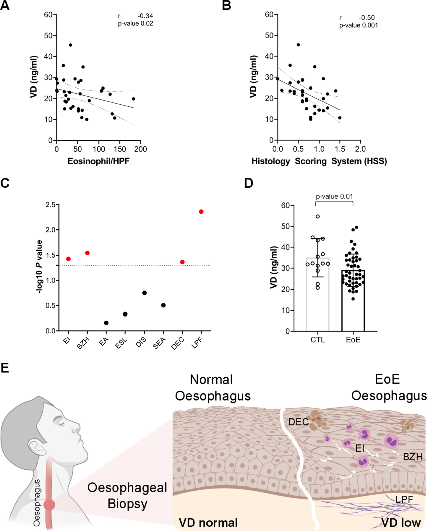
Role of vitamin D (VD) in eosinophilic oesophagitis. (A) Correlation of oesophageal eosinophilia with serum VD levels. (B) Correlation of EoE total Histology Scoring System (HSS) values with serum VD levels. (C) Significance (-log p value) of correlations of serum VD levels with individual HSS scores. Significant correlation in red. (D) Serum VD levels in patients with active EoE and controls (CTL). (E) Model of the oesophageal epithelium in EoE and control individuals. VD levels confer the disease state in which low VD levels induce EI, DEC, BZH and LPF (Created by BioRender). (A–D) Each data point is a mean of a technical duplicate ±SD of individual measurements. Statistics by unpaired one-tailed t-test (A) and Spearman correlation (B–D) for given p values (A–D) and Spearman correlation coefficients (r; A, B). BZH, basal zone hyperplasia; DEC, dyskeratotic epithelial cells; DIS, dilated intercellular spaces; EA, eosinophil abscess; EI, eosinophilic inflammation; EoE, eosinophilic oesophagitis; ESL, eosinophil surface layering; LPF, lamina propria fibrosis; SEA, surface epithelial alteration.