Fig. 1.

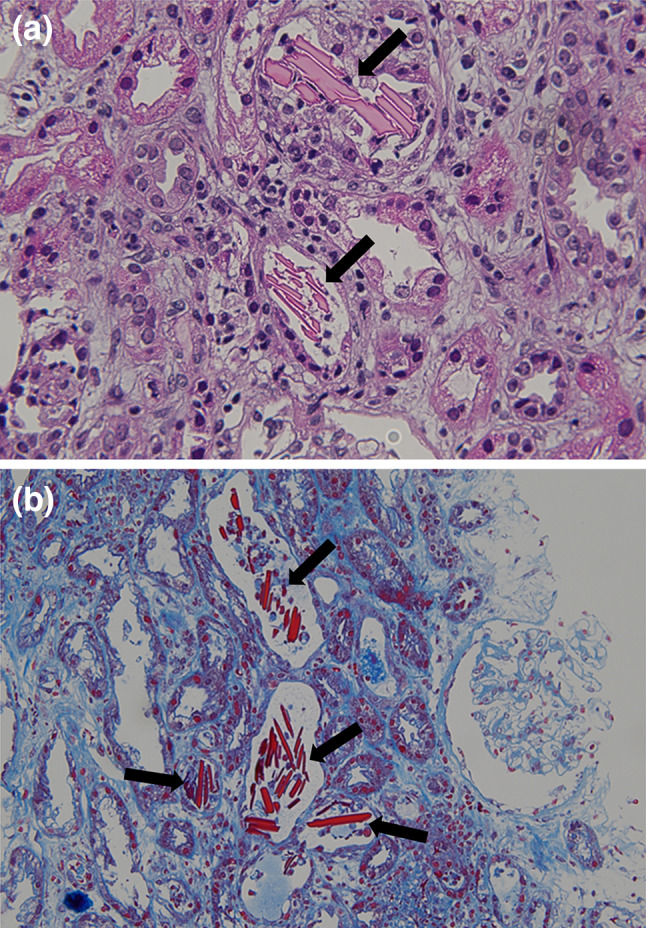
Light microscopy of the kidney showed crystalline cast filling the tubular lumens (arrow), injured tubular cells, and inflammatory cells infiltration of interstitium. (a Hematoxylin–eosin, × 400. b Masson trichrome, × 200.)

Light microscopy of the kidney showed crystalline cast filling the tubular lumens (arrow), injured tubular cells, and inflammatory cells infiltration of interstitium. (a Hematoxylin–eosin, × 400. b Masson trichrome, × 200.)