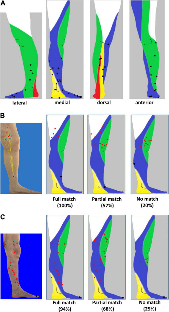
FIGURE 2.

Distribution of primary melanoma and in-transit metastases localized to the lower leg and foot. (A) Lateral, medial, anterior, and dorsal projection of primary melanoma (black) of 58 patients based on photographic images and mapping to distinct anatomic lymphatic drainages regions of leg: posteromedial (yellow), anteromedial (blue), anterolateral (green), and posterolateral (red). (B) Melanoma of the lower leg: three-dimensional (3D) image of a primary melanoma and corresponding in-transit metastases as well as the supposed lymphatic drainage (left image). Representative examples of individual patients with primary melanoma (black) on the lower leg with a full match of 8/8 (100%) of in-transit metastases (red) developing in the corresponding anteromedial lymphatic region (second image from the left) and metastases developing partially (8/14, 57%, second image from the right) or not predominantly within (2/10, 20%) the expected localization (right image). (C) Melanoma on the foot. Annotation of in-transit metastases (red) on a 3D photographic image (left image). Examples of representative individual patients with primary melanoma (black) and corresponding in-transit metastases (red) occurring fully, partially, or not (images second from the left, second from the right, and right) in the expected lymphatic drainage area.
