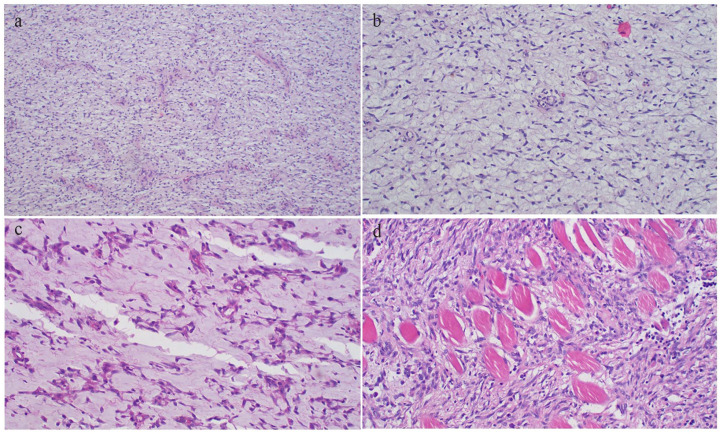
Figure 6.
(a) CMF demonstrating diffuse population of mononuclear spindle cells and admixed multinucleated giant cells (H&E 40× original magnification). (b) CMF highlighting the presence of lobules of stellate cells with variably myxoid to chondroid stroma, representing various stages of cartilaginous development (H&E 100× original magnification). (c) Photomicrograph of CMF showing cells having variable pink cytoplasm, bipolar to multipolar cytoplasmic extensions and oval to spindled nuclei with myxoid stroma (H&E 200× original magnification). (d) Infiltration of skeletal muscle by the tumor cells with moderate nuclear pleomorphism (H&E 200× original magnification).

