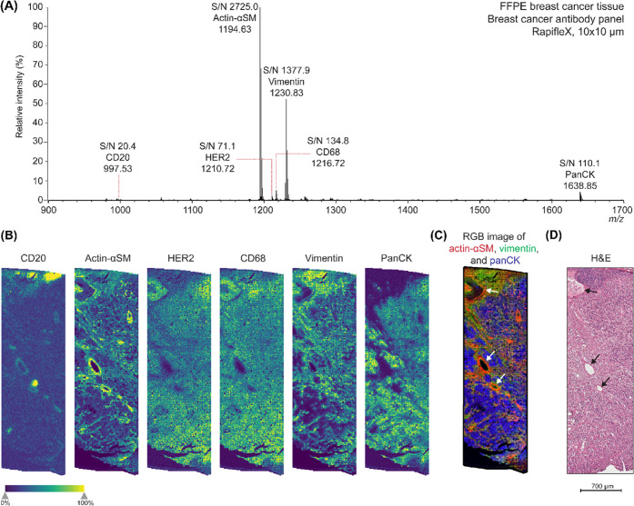
Figure 2.
Multiplex MALDI-IHC on the rapifleX for six biomarkers in FFPE human breast cancer tissue (patient 1). (A) Average spectrum of the MALDI-IHC measurement with the breast cancer antibody panel. All six peptide mass reporters were detected. The spectrum was TIC normalized and baseline-corrected. (B) Single-ion images of the mass reporters detected, which were CD20, actin-αSM, HER2, CD68, vimentin, and panCK, respectively. (C) RGB image of actin-αSM (red), vimentin (green), and panCK (blue). (D) H&E image of a consecutive section, showing similar structures as imaged with the MALDI-IHC. White (C) and black (D) arrows point out the vascular lining of blood vessels in the tissue section, which is also highlighted by actin-αSM (B, C).

