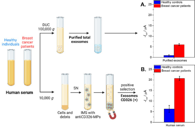
Figure 4.

Panel A shows the control of the purified total exosome population obtained by ultracentrifugation (100,000 × g) normalized according to the protein content (0.33 μg per assay). Panel B. Electrochemical genosensing of CD326+ exosomes from 1 mL of cell-free undiluted human serum (centrifuged at 10,000 × g) based on immunomagnetic separation with antiCD326-MP and further GAPDH transcripts detection. The whole procedure is also shown in Figure 1. In all cases, serum-derived exosomes from healthy controls (n = 10, pooled) and breast cancer (n = 10, pooled) patients were processed. The error bars show the standard deviation for n = 3. The raw amperograms are also shown in Figure S4 (Supplementary Data). Created with BioRender.com.
