
Figure 5.
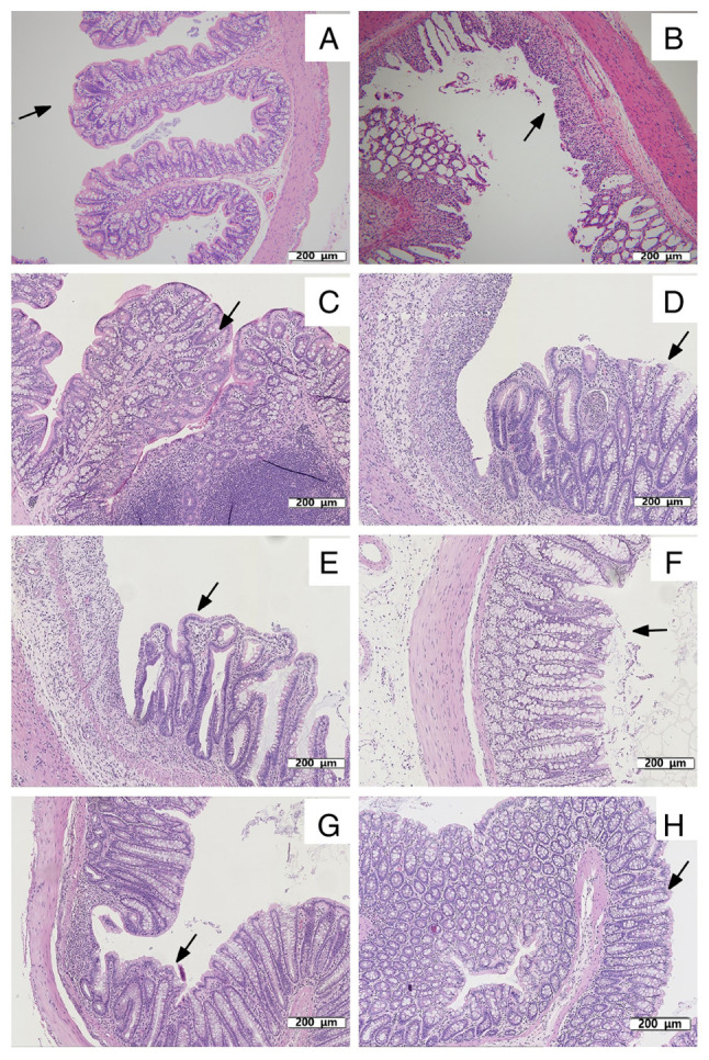
Histopathological examination of colon tissues of rats. (A) Colon tissue of rats in G1 on day 8. The mucosa is intact and free of inflammatory cell infiltration, as indicated by the arrow. (B) Acute colitis induced by DSS on day 8. Mucosal injury was shown as focal ulceration, epithelial necrosis and infiltration of inflammatory cells, as indicated by the arrow. Colon tissue of rats in (C) G1, (D) G2, (E) G3, (F) G4 (G) G5b and (H) G6 on day 15. (C) Colon tissue of rats in G1 on day 15. Intact mucosa and no inflammatory cell infiltration was indicated by arrows. (D) Colon tissue of rats in G2 on day 15. The mucosal injury was characterized by focal ulceration, epithelial necrosis and infiltration of inflammatory cells, as indicated by arrows. (E) Colon tissue of rats in G3 treated with prednisolone on day 15. Colon tissue exhibited a relatively intact mucosa with only a small infiltration of inflammatory cells, as indicated by arrows. (F) Colon tissue of rats in G4 treated with vitamin D on day 15. The colonic mucosa was intact but with some epithelial cell necrosis and infiltration of inflammatory cells, as indicated by the arrows. (G) Colon tissue of rats in G5b treated with vitamin E on day 15. The colonic mucosa was intact and infiltrated by a few inflammatory cells, as indicated by the arrows. (H) Colon tissue of rats in G6 treated with vitamin D and vitamin E on day 15. The colonic tissue mucosa was more intact than that in G4 and G5b, as indicated by the arrows. Treatment with vitamin D, vitamin E, vitamin D + vitamin E and prednisolone reduced the morphological alterations associated with DSS administration and protected the mucosal architecture. Colon tissues in the figures were all stained with hematoxylin and eosin. Scale bar, 200 µm; magnification, x110. G1, control group; G2, 5% DSS group; G3, prednisolone group; G4, vitamin D group; G5a-c, vitamin E (low, medium and high) groups; G6, vitamin D + vitamin E group; DSS, dextran sodium sulfate.