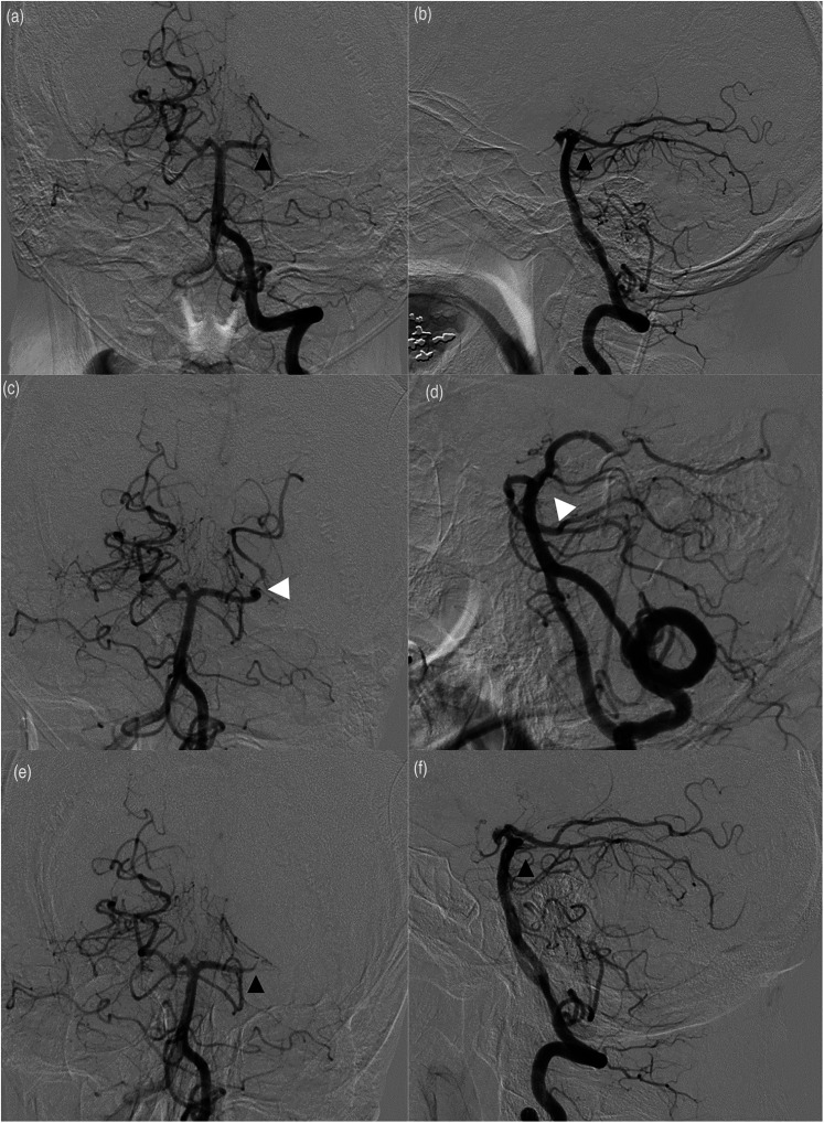
Figure 1.
(a) anteroposterior (AP) and lateral (b) views of a left vertebral artery selective catheterization demonstrating an occlusion of the left posterior cerebral artery (PCA) at the P1-P2 junction (black arrows). After stent retriever-assisted aspiration, partial reopening of the PCA (white arrows) was witnessed, as shown in the AP (c) and lateral (d) views. However, despite administering eptifibatide, further imaging demonstrated reocclusion of the vessels (E, AP; F, lateral) (black arrows).

