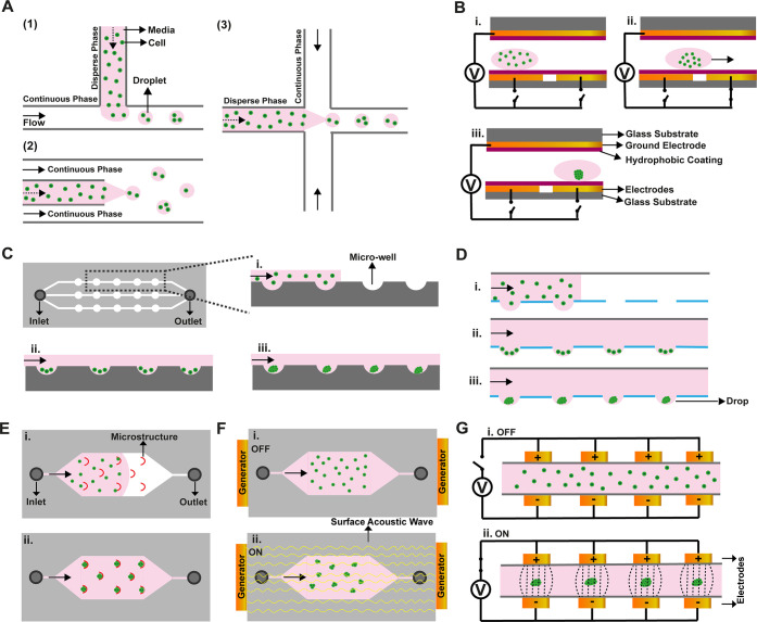
Figure 2.
Illustrations for microfluidic-based methods in spheroid formation. (A) Droplet-based methods: (1) T-junction breaks up the dispersed phase with a sheath flow; (2) in co-flowing, the dispersed phase is generated with a needle or tube; and (3) flow focusing uses two sheath flows for breaking of the dispersed phase. (B) Electrowetting generated with an electrode pattern. (C) Microwells in channels. (D) Microfluidic hanging drop with open wells in the channel. (E) Microstructure for cell trapping. (F) Acoustic manipulations. (G) Dielectrophoresis with a nonuniform electrical field. (i), (ii), and (iii) represent spheroids formation steps in all images.

