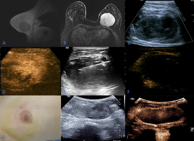
Figure 1.
A 37-year-old female with huge mass in the left breast; (A, B) The location of the tumor on enhanced MRI; (C); Sonographic appearance of the tumor on 2D-Ultrasound; (D) Contrast-enhanced ultrasound showed homogeneous hyperenhancement of the tumor during the arterial phase; (E) During ultrasound-guided microwave ablation; (F) The area of ablation showed no enhancement in arterial phase and venous phase after ablation; (G); The skin of the breast was slightly ecchymosis after ablation, and the appearance of the breast was intact; (H, I) After 10 months of follow-up, two-dimensional ultrasound showed that the mass was significantly reduced, and CEUS showed that there was no enhancement in the arterial phase and venous phase of the ablation area.

