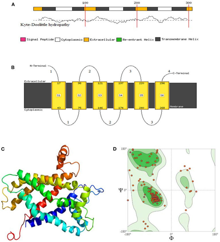
Figure 6.
(A) Membrane helix prediction with support vector machines with Kyte-Doolittle hydropathy plot. (B) Schematic figure of predicted extracellular, cytoplasmic, and transmembrane helices. (C) Top 3D model of avian UCP [Model dimensions (Å): X: 53.347, Y: 74.255, Z: 50.565, Image colored by the rainbow N → C terminus]. (D) Ramachandran plot with 88.46% favored. A dihedral angle of a protein is the internal angle of polypeptide backbone at which two adjacent planes meet. The conformation of the backbone can be described by two dihedral angles per residue, because the backbone residing between two juxtaposing Cα atoms are all in a single plane. These angles are called ϕ (phi) which involves the backbone atoms C-N-Cα-C, and ψ (psi) which involves the backbone atoms N-Cα-C-N.

