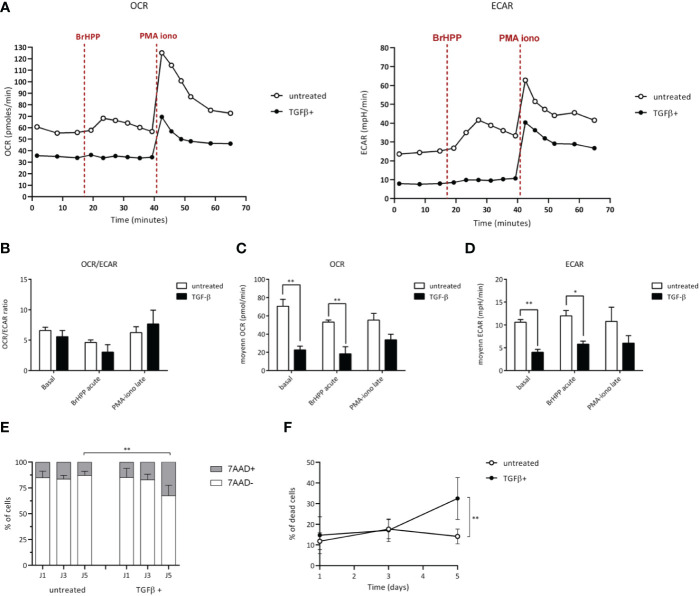
Figure 4.
TGF-β alters the metabolism of Vγ9Vδ2 T cells. Vγ9Vδ2 T cells were treated (closed symbols), or not (open symbols), with TGF-β (10 ng/mL) for 72 h. Metabolic activities of T lymphocytes were measured using Seahorse XFp. (A-D) Monitoring of mitochondrial oxygen consumption rate (OCR) and extracellular acidification rate (ECAR) were performed at resting state, after BrHPP stimulation or PMA-ionomycin injection as described in the Materials and Methods section. (A) Representative experiments. Rates were calculated as follows: difference between the mean of the 3 values in the absence of substrate and the mean of the 4 values after injection of the substrate, as well as a ratio of the 2 for each condition (B-D). (E) and (F) Cell death induced by TGF-β added within the media of Vγ9Vδ2 T cells (10 ng/mL for 72 h) was measured by measuring fluorescent non-viable cells following 7-AAD staining after the indicated culture timepoints days of culture. n=6, mean ± SD. Mann-Whitney *p<0.05; **p<0.01. PMA-iono: PMA+ionomycin (positive control).

