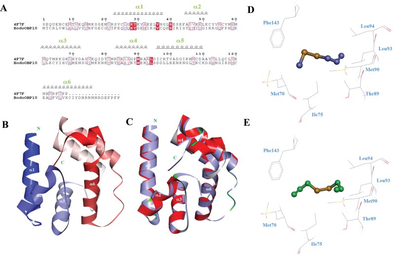
Fig. 4.
3D structural modeling and molecular docking of BodoOBP10. (A) The sequence alignment between the template protein AgamOBP20 and the target protein BodoOBP10; the black coil represents the α-helix; (B) The 3D model of the target protein BodoOBP10 based on the crystal structure of the template protein of AgamOBP20; the position of α-helix is marked by white; (C) The alignment plot of the target protein BodoOBP10 (red) and the template protein AgamOBP20 (purple, ID: 4F7F). Predicted binding mode and key residues between the target protein BodoOBP10 and methyl allyl disulfide (purple) (D), diallyl disulfide (green) (E). Residues are marked in blue.

