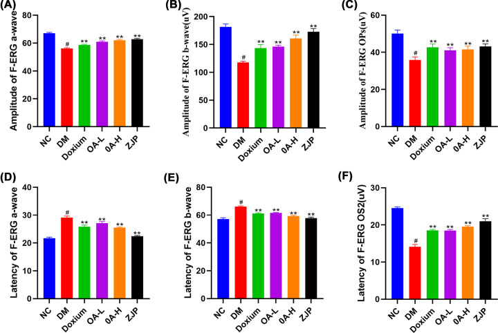
Figure 5. Retinal electrophysiological examination.
Panels (A–C) are the a-wave, b-wave and Ops amplitude of F-ERG;panel (D–F) represent the a-wave, b-wave and OS2 latency of F-ERG. All data were expressed as the mean ± SD (n=3). Compared with the normal group, #P<0.01; Compared with the model group, **P<0.01.

