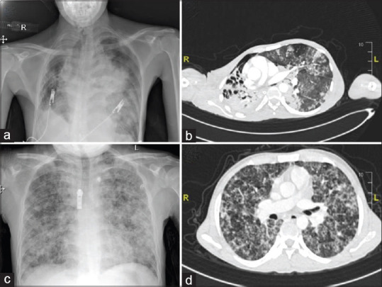
Figure 1.

(a) The chest roentgenogram of patient 1 shows decreased right lung volume with cavitary lesions in upper and mid zones, and the left mid zone shows a small area of sub-segmental consolidation with preserved lung volume suggestive of TB and COVID. (b) Computed tomography of the thorax of the same patient [patient 1] shows shifting of the upper mediastinum towards the right side with multiple cavitary lesions in the right upper lobe. A few atelectatic bands were noted in the left upper lobe and left lower lobe with sub-segmental collapse. (c) Chest roentgenogram of patient 2 shows areas of reticulonodular opacities in the bilateral lung parenchyma (left > right) with a few areas of fluffy opacities suggestive of military tuberculosis. (d) Corresponding computed tomography of the thorax of patient 2 shows the thickening of interlobular septa and small nodules. Ground glass opacities were seen in the lower lobe (image not in set)
