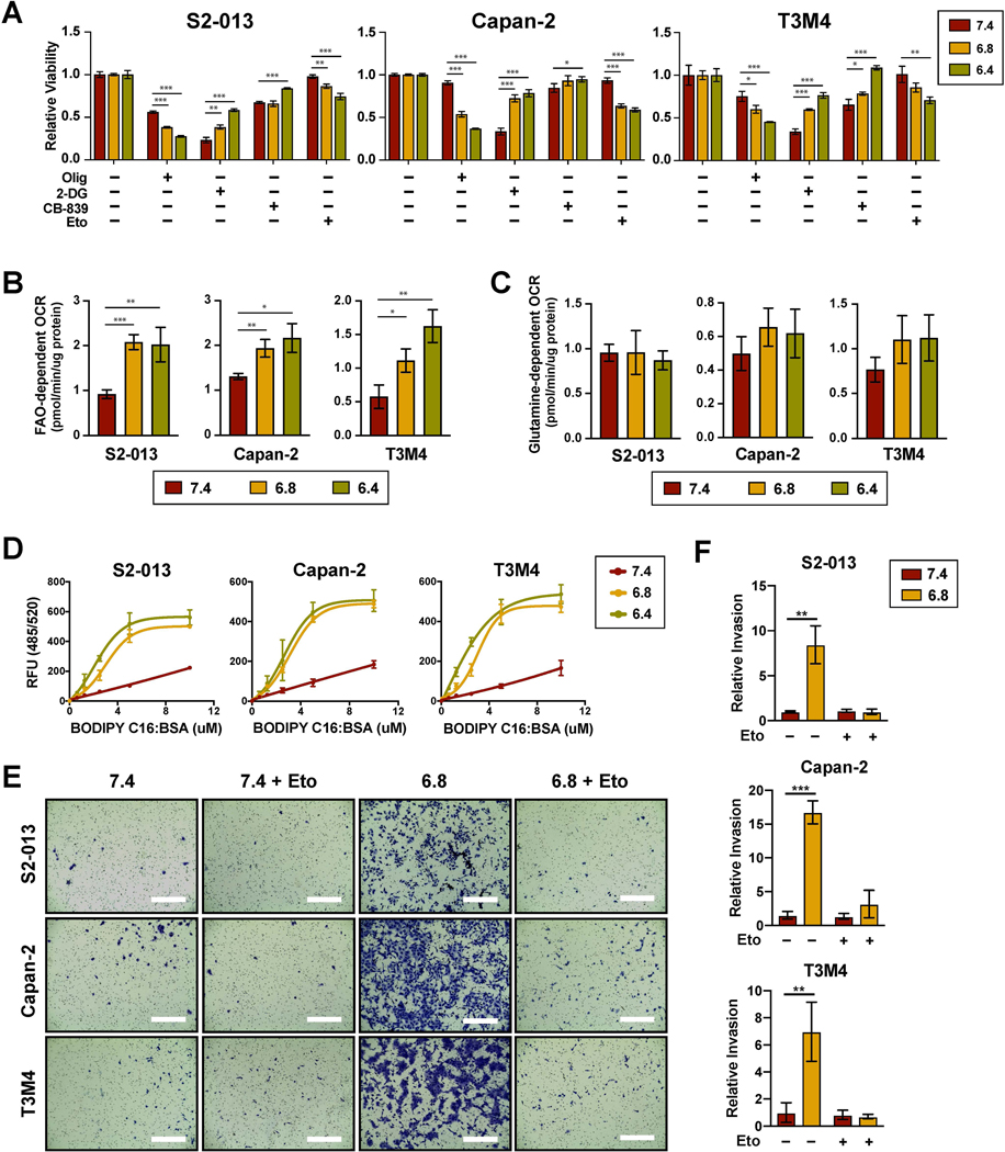
Figure 3. Low pHe increases PDAC dependency on fatty acid oxidation.
(A) Relative cell viability following 48-hour treatment with the indicated metabolic inhibitors compared to untreated controls (n=3). FAO and glutamine-dependent OCR as determined by the decrease in basal OCR following injection of etomoxir (B) (n=3) or CB-839 (C) (n=3), respectively. (D) Lipid uptake measured by incubating cells with fluorescently labeled palmitate (BODIPY C16) conjugated to fatty acid-free BSA for 15 min following 24-hour pre-culturing under the indicated pHe conditions (n=3). (C) Representative images from Matrigel invasion assays. Bar, 400μm. (D) Quantification of relative invasion (n=2). Data are shown as mean ± SD. *p < 0.05; **p < 0.01; ***p < 0.001.

