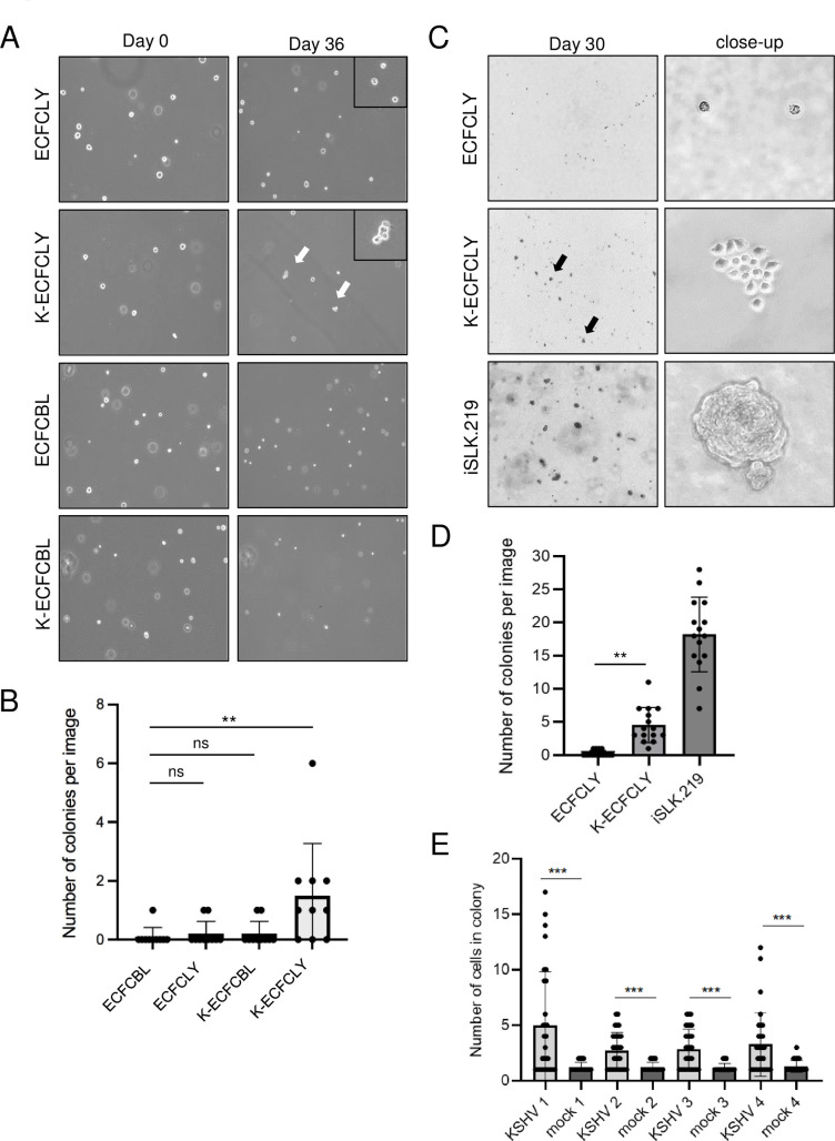
Fig 3. KSHV confers a survival advantage to lymphatic ECFCs grown in soft agar.
A. Single cells suspensions of mock or wtKSHV-infected lymphatic and blood ECFCs were embedded 48 h.p.i in soft agar overlaid with 0.5ml growth media, and media was replenished every 2–4 days. Images were taken 36 days post plating. B. The number of colonies with two or more cells at 36 days post plating were quantified. These experiments were performed up to eight times with similar results. C. Lymphatic ECFCs mock- or rKSHV.219-infected were embedded 5 d.p.i. in soft agar. Stably KSHV-infected iSLK.219 was used as a positive control cell line. Images were taken at 30 days post plating. D. Quantification of the number of colonies consisting of at least 3 cells from the samples in C. E. Quantification of the number of colonies at 30 days post plating from all four different ECFCLY donors infected with rKSHV.219 and embedded 5 d.p.i. in soft agar. Experiments were performed at least three times with similar results. ** p < 0.01, *** p < 0.001.

