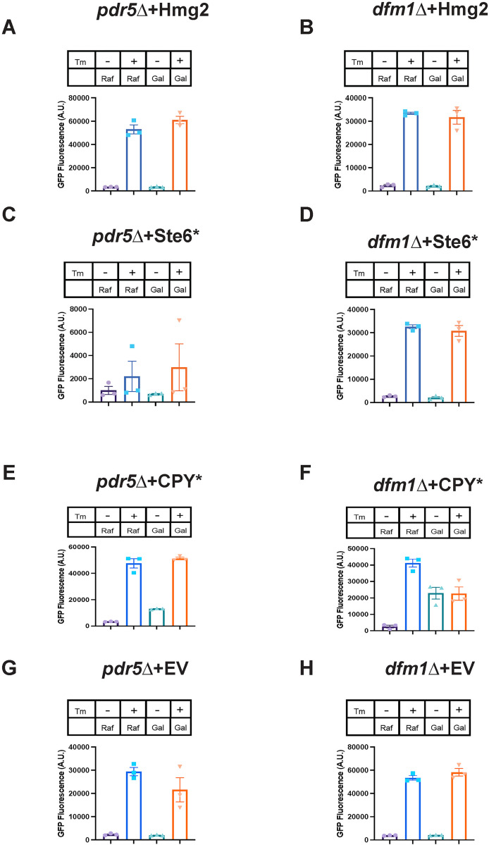
Fig 5. Misfolded membrane protein stress in dfm1Δ cells does not activate the UPR.
(A) UPR activation for indicated strains with overexpression of a misfolded integral membrane protein. pdr5Δ cells containing GALpr-Hmg2-6Myc and 4xUPRE-GFP (a reporter that expresses GFP with activation of the UPR) were measured for GFP expression using flow cytometry every hour for 5 hours starting at the point of galactose induction and tunicamycin or equivalent volume of DMSO was added at the 1-hour time point. Figure depicts the GFP fluorescence in A.U. for indicated conditions 5 hours post-galactose addition. In figure legend, “Gal” indicates addition of 0.2% galactose to cultures and “Raf” indicates addition of 0.2% raffinose to culture, and “Tm” indicates presence (+) or absence (-) of 2 μg/mL tunicamycin. (B) Flow cytometry-based UPR activation assay as described in (A) except using dfm1Δ cells. (C, E, and G) Flow cytometry-based UPR activation assay as described in (A) except using cells containing GALpr-Ste6*-GFP, GALpr-CPY*-HA, or EV, respectively. (D, F, and H) Flow cytometry-based UPR activation assay as described in (B) except using cells containing GALpr-Ste6*-GFP, GALpr-CPY*-HA, or EV, respectively. Data information: All data are measured mean ± SEM; N = 3 biological replicates. The data underlying this figure can be found in Table A–H in S1 Data (Sheet 1). A.U., arbitrary unit; EV, empty vector; UPR, unfolded protein response.

