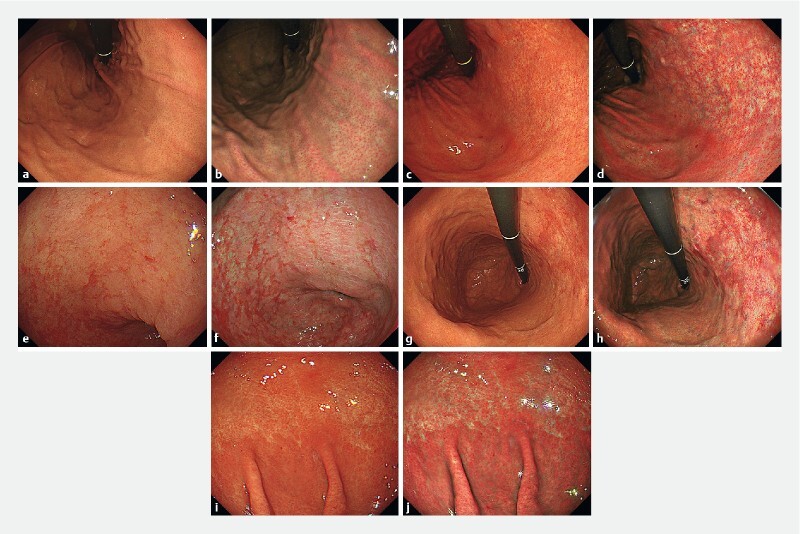
Fig. 1 .

Representative endoscopic images. Endoscopic images of the regular arrangement of collecting venules (RAC) in the stomach. a WLI. Venular accumulation is noted with an increase in minute points, and it is regularly distributed in the body of the stomach. b TXI. Regular tiny veins are depicted more clearly. Endoscopic images of the atrophic border in the stomach. c WLI. An atrophic border is found at the lesser curvature of the gastric body. d TXI. The atrophic border is more identified. Endoscopic images of intestinal metaplasia in the stomach. e WLI. Slightly elevated whitish patches are observed in the antrum of the stomach. f TXI. Whitish patches become more noticeable. Endoscopic images of map-like redness in the stomach. g WLI. Reddish depressed area is observed in the atrophic area of the gastric body. h TXI. The reddish depressed area appears more vivid. Endoscopic images of diffuse redness in the stomach. i WLI. A uniform redness is observed on the non-atrophic mucosa of the fundic gland in the gastric body. j TXI. Visualization of the reddish area is enhanced.
