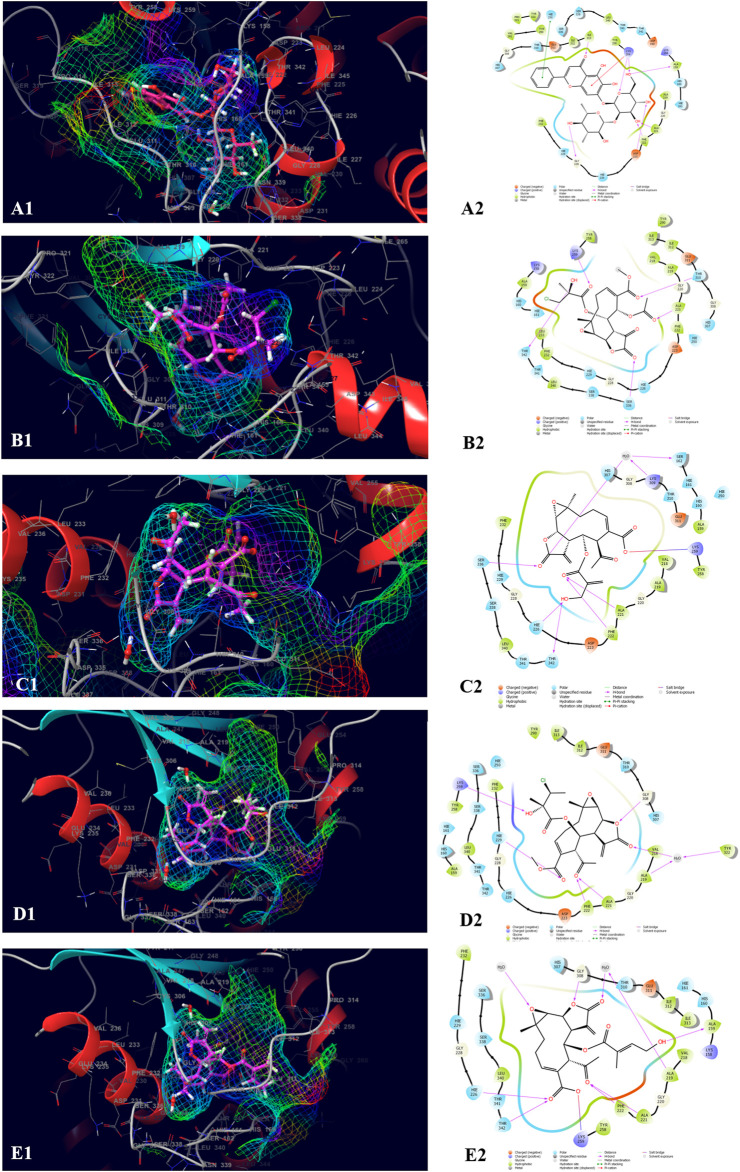
FIGURE 4.
Molecular docking interactions of metabolites in the binding pocket of PDB ID: 3ELB. [3D and 2D representation of Baicalein 7-O-diglucoside (A1,A2), 8-Deepoxyangeloyl-8-2-hydroxy-3-chloro- isobutyroyl-enhydrin (B1,B2), 8-Desacylenhydrin-4-hydroxymethacrylate (C1,C2), 8-Deepoxyangeloyl- 8-chloro- 2-hydroxy-2-methylbutyroylenhydrin (D1,D2) and 8-Desacyl enhydrin-4-hydroxytiglate (E1,E2) in the binding pocket of (PDB ID: 3ELB)].

