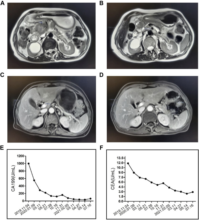
FIGURE 2.
Contrast-enhanced Magnetic Resonance Imaging (MRI) of the upper abdomen showed the patient’s pancreatic carcinoma before (A) and after (B) the treatment of PD-1, HIFU and Gemcitabine. MRI showed the patient’s liver metastasis before (C) and after (D) the treatment. CA199 (E) and carcinoembryonic antigen (CEA) (F) index of the patient.

