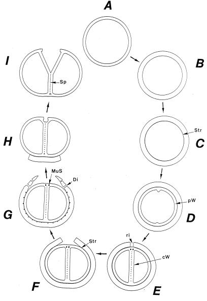
FIG. 17.
Wall regeneration sequences after chloramphenicol treatment. Growth-inhibiting bacteriostatic antibiotics, such as chloramphenicol, do not interfere with the synthesis of wall material. Consequently, under such bacteriostatic conditions the slowly growing staphylococci are capable of forming huge masses of wall material, resulting in extremely thick cell walls. After being transferred to normal growth medium, a comprehensive sequence of cutting and separation processes in the thick wall material is initiated to restore the normal width of the cell wall. This simplified schematic drawing gives a survey of these processes. (A to B) Wall thickening. By chloramphenicol-mediated, successive underlayerings of the peripheral cell wall with newly synthesized wall material, the staphylococcal cell wall becomes considerably wider. Reference figure, Fig. 16a. (C) Formation of a stripping layer. After transfer into normal growth medium, staphylococci initiate wall regeneration by placing a so-called stripping layer (Str) beneath the thick peripheral wall. Reference figure, Fig. 16b. (D) Synthesis of a new peripheral wall. Under the stripping layer a completely new peripheral cell wall (pW) is synthesized via inside-to-outside processes of wall assembly. Reference figure, Fig. 16c. (E) Cutting through of the old peripheral wall. Removal of the old and apparently obstructive peripheral wall is initiated by an asymmetrical cutting through of this wall via forming a so-called cutting rim (ri) above the cross wall (cW) which is now synthesized. Reference figure, Fig. 16c. (F) Onset of sequential wall-stripping processes. By activation of autolytic wall hydrolases within the stripping layer (Str), the old superfluous peripheral wall is sequentially peeled off from the underlayered new peripheral wall. Reference figure, Fig. 16d and e. (G) Disintegration of stripped cell walls. The detached old cell wall is disintegrated either already during the stripping of the old peripheral cell wall or after this process. The disintegrating processes start from periodically arranged holes (disintegration system [Di]) located between the stripping layer and the old peripheral wall and result in the liberation of rather large pieces of the old, peripheral cell wall. Reference figure, Fig. 16f. MuS, murosomes. (H and I) Normal growth after detachment of the old cell wall. As soon as the old peripheral wall formed under chloramphenicol is removed, normal growth takes place, starting in staphylococci with completed cross walls, by murosome-mediated punching of pores into the new peripheral wall and subsequent cell separation. Reference figures, Fig. 10a and 13a. Sp, splitting system.

