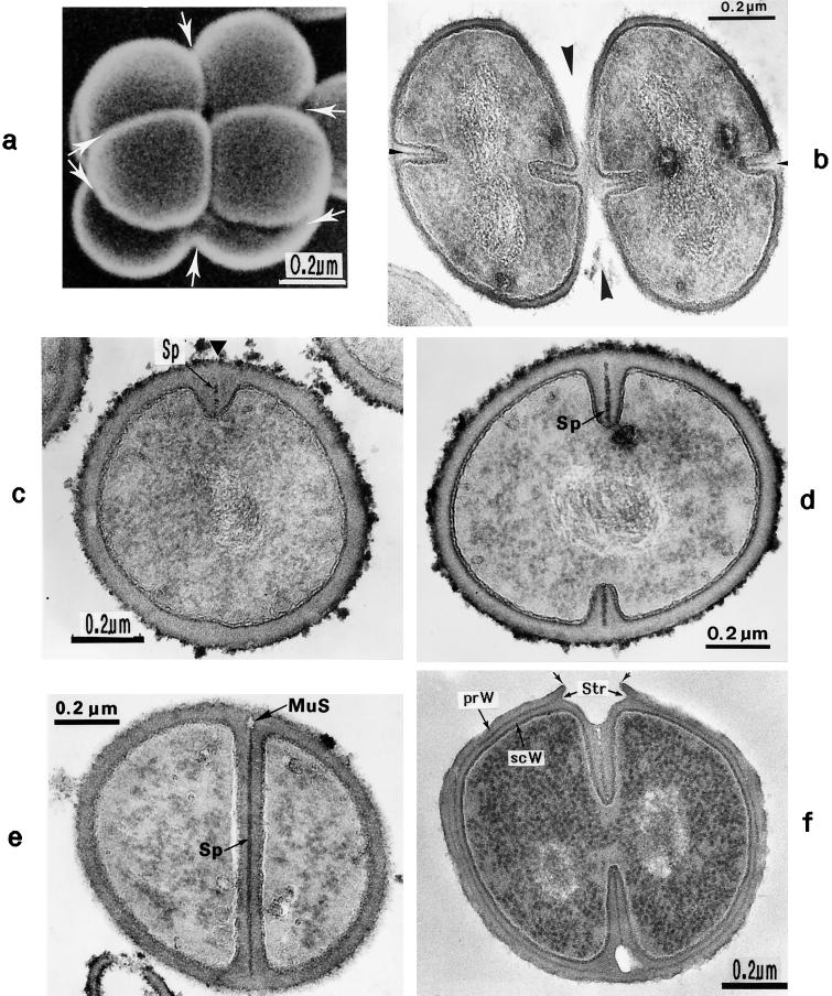
FIG. 2.
Scanning electron micrograph (a) and thin sections of staphylococcal cells (b to f). (a) A packet of eight staphylococcal cells was induced by liquoid; this packet is derived from one bacterium by three consecutive cell divisions, each having changed its direction at an angle of 90° to the preceding division plane. The three division planes are indicated by arrows (reproduced with permission from reference 139). (b) Characteristic alternation of consecutive division planes (arrowheads) (reproduced with permission from reference 41). (c) Asymmetrical initiation of cross wall formation (arrowhead), Sp, splitting system (reproduced with permission from reference 41). (d) Centripetal growth of the closing cross wall. The splitting system (Sp) appears as a darker central cross wall layer. (e) At the cell periphery, above the closed cross wall with its splitting system (Sp), there is one of the murosomes (MuS) (reproduced with permission from reference 50). (f) After a 2-h exposure to the antibiotic batumin (1 μg/ml) the peripheral wall appears to be differentiated into an outer layer, the so-called primary wall (prW), and an inner layer, the so-called secondary wall (scW). The dark line between these two layers represents the so-called stripping system (Str) of the staphylococcal cell wall which is involved in cell wall turnover. The remnants of the cutting through of the primary wall, the so-called clefts, are marked by arrows.

