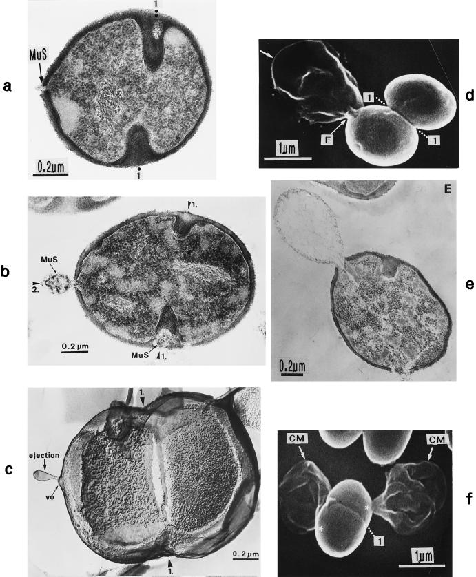
FIG. 20.
Thin sectioning (a, b, e), freeze fracturing (c) and scanning electron micrographs (d, f) of staphylococcal cells grown in the presence of 0.1 (a, b, e) or 10 (c, d, f) μg of penicillin/ml. Some cultures were grown in medium supplemented with 3% NaCl and were subsequently transferred to medium with low osmolarity (d and f). (a) The murosome (MuS) has, finally, perforated the outer layers of the peripheral cell wall, the so-called primary wall, at the initiation site of the second division plane. 1, first division plane (reproduced with permission from reference 50). (b) After perforation of the outer, primary wall at the site of the second division plane (2), the murosome (MuS) is ejected together with some plasmatic material (arrowhead). 1, first division plane (reproduced with permission from reference 48). (c) Even in the presence of high concentrations of penicillin the ejection of the murosome can be detected at the initiation site of the second division plane. This ejection starts from a local bulge of the peripheral wall (vo). 1, first division plane. (d) At high concentrations of penicillin the explosion-like liberation of the cytoplasmic membrane (short arrow) has caused a considerable enlargement of the small, murosome-induced wall perforation (E) at the second division plane. 1, first division plane (reproduced with permission from reference 53). (e) A similar explosion-like liberation of the cytoplasmic membrane occurs at low penicillin doses (reproduced with permission from reference 48). (f) At high concentrations of penicillin (10 μg/ml) murosome-induced explosions (stars) can take place simultaneously in the second division planes of both daughter cells, liberating at the same time two cytoplasmic membranes (CM). 1, first division plane.

