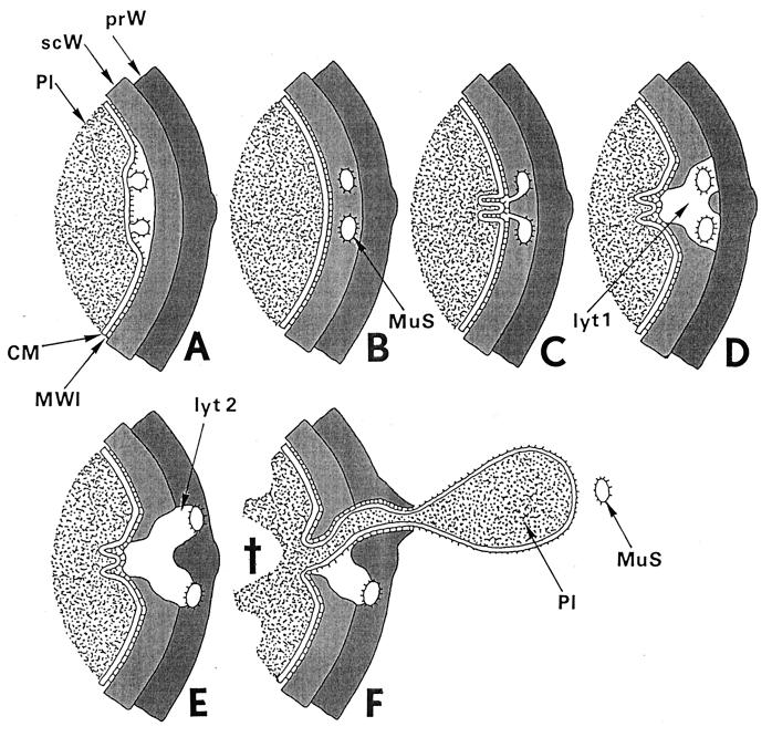
FIG. 22.
Fatal effects of “normal” lytic wall processes. Two consecutive lytic processes of wall morphogenesis in control cells, induced by the same murosomes in an interval of 10 min, are the key events for understanding penicillin-induced death: (i) initiation of cross wall formation and (ii) initiation of cell separation. In penicillin-treated staphylococci both processes take place during the same phase of wall morphogenesis, at the same site, and in the same way as in control cells. Only some penicillin-induced variations in the distribution of wall material proved to be fatal (for an overview, see Fig. 21). The schematic drawing seeks to provide a visual aid for a better understanding of these crucial processes. The depicted area is the site where both these lytic processes take place during the second cell cycle after the addition of penicillin. (A to D) Initiation of cross wall formation. (A) At the site of the second division plane, murosomes are formed by the cytoplasmic membrane (CM) or its membrane-wall interlayer (MWI) via an evagination process. Pl, cytoplasm; prW, primary wall; scW, secondary wall. Reference figures, Fig. 19b and 6f to h. (B) Immediately before cross wall formation starts, the murosomes (MuS) are found to be located in the lower layer of the peripheral cell wall, the so-called secondary wall. Probably, they have penetrated into this secondary wall or they are formed together with this wall layer. Reference figures, Fig. 19b and c and 6c (see also Fig. 8). (C) Lytic processes of the murosomes, directed to the center of the cell, separate the secondary wall into three parts. Folds of the cytoplasmic membrane indicate the first steps for cross wall formation. Reference figure, Fig. 6d and e. (D) The central part of the secondary cross wall starts the formation of the central, “transitory” layer of the future cross wall while the other parts initiate the “permanent” layers (see Fig. 7). However, while in control cells cross wall formation goes on until it is completed, in the presence of penicillin, cross wall formation at this site ceased because the necessary wall material is deposited at another site; furthermore, lytic processes (lyt 1) within the secondary wall proceed, leaving behind a disintegrated sector in the secondary wall. Reference figure, Fig. 6d and 19c to f. (E and F) Fatal initiation of cell separation. (E) In spite of the fact that in the presence of penicillin there is no cross wall material deposited in the second division plane, staphylococci start normal cell separation with murosome-mediated punching of pores into the primary layer of the peripheral wall via outward directed lytic processes (lyt 2). Reference figure, Fig. 20a. (F) As soon as one of the murosomes (MuS) has succeeded in perforating the outer layer of the peripheral wall and is released into the growth medium, the cell will burst and eject limited amounts of its cytoplasm (Pl), due to its extremely high internal turgor. This death (cross) happens only because a protecting cross wall is missing beneath the single wall perforation. Reference figure, Fig. 20 b to f.

