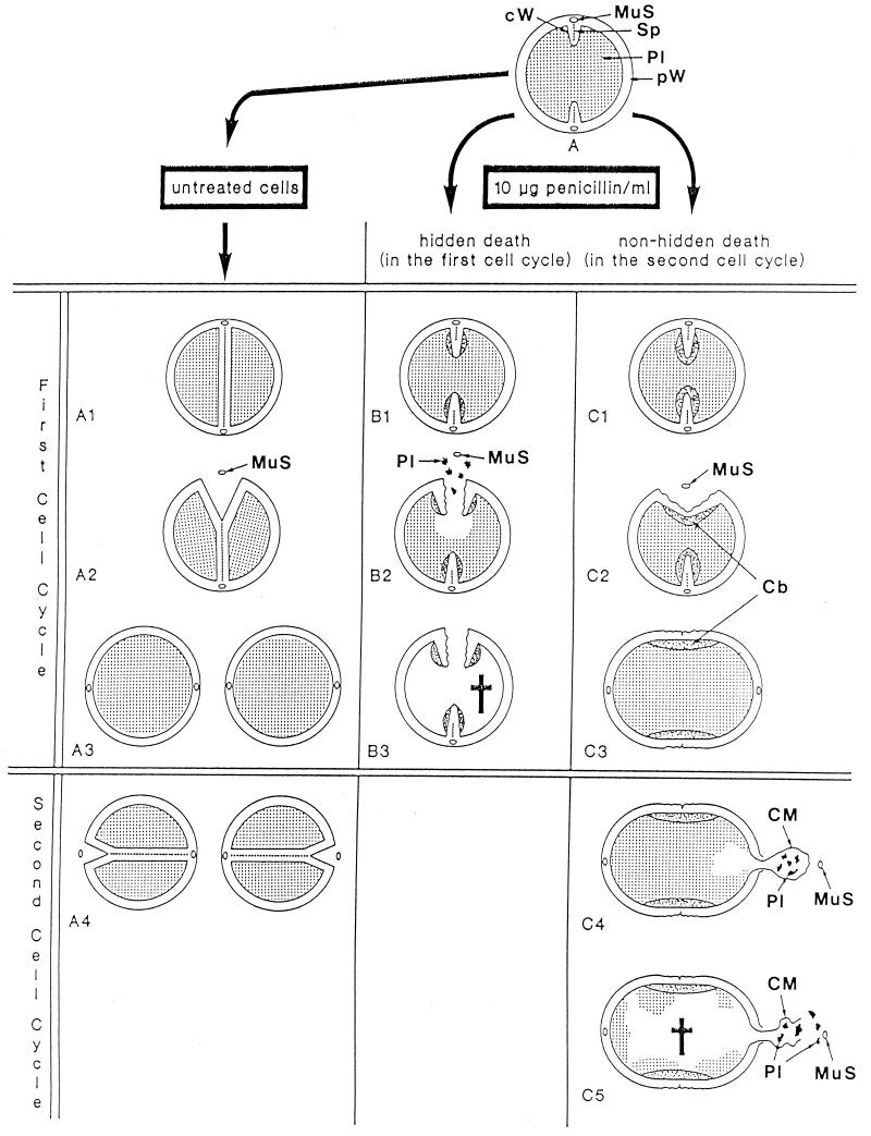
FIG. 25.
Hidden death at high penicillin concentrations. Schematic drawing illustrating the fate of staphylococcal control cells (A1 to A4) and of cells in the presence of high penicillin concentrations (B1 to B3 and C1 to C5) during the first and the second cell cycles. (A1 to A3) Control cells during the first cell cycle. (A1) Untreated cells divide by completion of their cross wall; (A2) they initiate cell separation via murosome-induced punching of pores into the peripheral wall above the completed cross wall; (A3) two daughter cells have been generated by this first cell separation. MuS, Murosome. (B1 to B3) Hidden death during the first cell cycle. (B1) In the presence of high concentrations of penicillin the formation of the splitting system stops and cell wall synthesis is largely inhibited. Instead of compact, highly organized cross walls, only some loose fibrillar cross wall material is synthesized which is mainly deposited laterally on the ingrowing cross walls, leaving the tips of the cross wall unprotected. Reference figure, Fig. 24a. (B2). Like in untreated cells (A2), cell separation is then initiated in spite of the fact that cross wall formation has not yet been completed. After liberation of the murosomes (MuS), cell separation proceeds along the splitting system synthesized before the action of the drug. Because of the high internal pressure and since the tips of the cross wall are not sufficiently protected by fibrillar cross wall material, the affected cells erupt granular cytoplasm (Pl) through slit-like openings in the peripheral wall. Reference figure, Fig. 24d. (B3) The incomplete cross wall of the first division plane is ripped up along the splitting system, the cell loses considerable parts of its cytoplasm and dies, leaving behind only empty cell walls which still preserve the shape of the primary staphylococcus. Reference figure, Fig. 24c. (C1 to C5) Nonhidden death during the second cell cycle. (C1) For still unknown reasons, a certain percentage of staphylococci treated with high doses of penicillin is still capable of synthesizing considerable amounts of fibrillar cross wall material after blocking the formation of the splitting system. In those cases not only the lateral parts but also the tips of the ingrowing cross walls of the first cell cycle are covered with these fibrils. Reference figure, Fig. 24e. (C2) Since the cross wall fibrils at the tips are capable of welding with each other, initiation of cell separation via murosome-mediated perforations of the peripheral wall (MuS) and ripping up of the cross wall along the primary splitting system will not result in a loss of cytoplasm and in death, and this in spite of the fact that their cross walls are not completed either: a connecting bridge (Cb) formed by the welding of fibrillar cross wall material proves to be tough enough to resist the high internal pressure. Reference figure, Fig. 24e. (C3) Consequently, those cells capable of welding together the individual primary cross walls of the prospective daughter cells can survive at least the first division cycle in the form of more or less elongated cells. Reference figure, Fig. 24f. (C4) Like in normal cells (A4), cell separation during the second cell cycle may be initiated via murosome-mediated formation of peripheral pores in the second division plane (MuS). Since hardly any cross wall material is deposited at this site, the extremely high internal pressure widens one of these pores, and part of the cytoplasmic membrane (CM) together with cytoplasmic constituents (Pl) is thrown out via an explosion-like ejection. Reference figure, Fig. 20d. (C5) Rupture of the thrown-out part of the cytoplasmic membrane (CM) results in the release of much of the granular cytoplasm (Pl), and the cell dies. Reference figure, Fig. 20f. (Modified from reference 53.)

