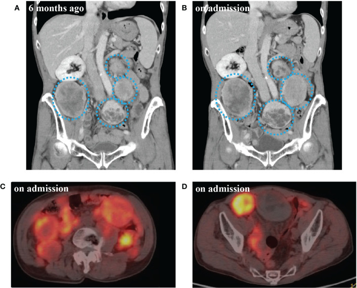
Figure 1.
(A) Computed tomography (CT) scan of abdomen 6 months before admission. (B) CT scan at the time of admission. (C) 68Ga-labelled 1,4,7,10-tetraazacyclododecane-N,N’,N’’,N’’’-tetraacetic acid-d-Phe1-Tyr3-octreotide positron emission tomography/CT (DOTATOC-PET/CT) at abdominal level on admission. (D) DOTATOC-PET/CT at pelvic level on admission. CT scan of multiple intra-abdominal tumors of increasingly large size (blue dotted circle). DOTATOC PET/CT showing uptake of radiotracer in the tumors.

