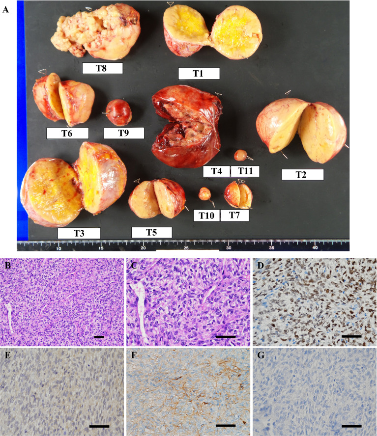
Figure 3.
(A) Macroscopic view of resected tumors. T1–11 represent tumor #1–11. (B–G) Microscopic findings of the representative resected tumor. (B, C) Hematoxylin and eosin staining showed that the tumor was composed of spindle to ovoid cells forming the patternless pattern. Immunohistochemical stains of tumor #10 show tumor cells to be positive for (D) signal transducer and activator of transcription 6 (STAT6), (E) IGF2, (F) somatostatin receptor 2 (SSTR2) and negative for (G) SSTR5. Scale bar: 50 μm.

