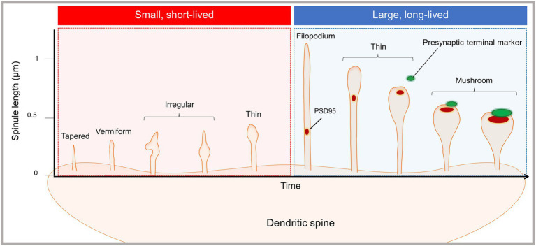
Figure 1.
Emergence of morphologically and functionally distinct spinule subsets. The proposed spinule classification system is based on our recent live-cell, time-lapse, enhanced resolution imaging study of spinules in cortical mouse neurons (Zaccard et al., 2020). The majority of spinules emerging from mushroom spines were dynamic and short-lived, existing for sec and up to 1 min, while a minority of spinules were larger and long-lived. Short-lived spinules were typically less than 0.5 microns in length, and displayed diverse morphologies, e.g., tapered, vermiform, irregular, and thin shapes. Long-lived spinules existed for ≥60 s, in some cases exceeding imaging durations up to 10 min, and were typically between 0.5 and 1 microns in length. Additionally, a subset of filopodia-shaped spinules extended up to several microns. Both filopodia and thin spinules often trafficked PSD fragments or contacted presynaptic terminals, but not both. Mushroom spinules were the most stable, with colocalized presynaptic and postsynaptic markers at enlarged spinule tips. Synaptic activity enhanced the number of spinules and the proportion of elongated, long-lived spinules.

