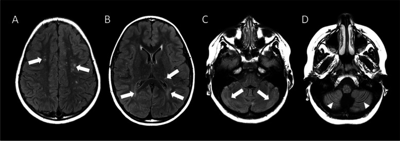
Fig. 5.
Axial FLAIR images obtained 3 months after hospitalization. A-D. Sequential FLAIR MRI from superior (A) to inferior (D) demonstrate small hyperintense lesions within the centrum semiovale (arrows; A) and periventricular white matter and left thalamus (arrows; B) that are more numerous than on the previous examination. Signal abnormality in the basal ganglia and thalamus is substantially improved. Abnormal hyperintense signal presumably representing gliosis (arrows; C) and fissural prominence denoting volume loss (arrowheads; D) are seen in the cerebellum

