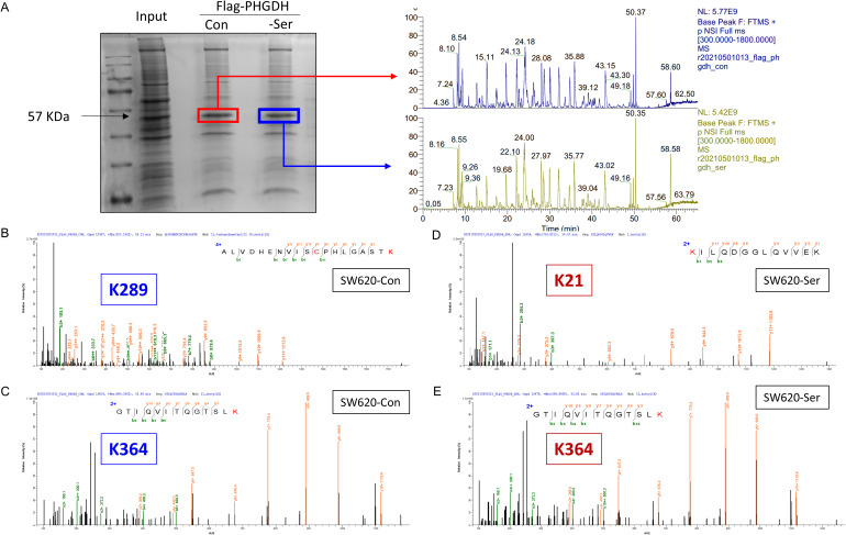
Figure 5.
Post-translational modification changes of PHGDH protein under serine deprivation. (A) 293T cells transfected with Flag-PHGDH plasmid in complete medium and serine withdrawal medium, respectively, and the protein samples collected after Immunoprecipitation were electrophoresed by SDS-PAGE and the protein distribution maps stained with Kaomasbrilliant blue. The target protein (∼57KD) was collected and analyzed by HPLC-MS. The figure on the right shows the primary mass spectrum of the target protein. (B) and (C) Under complete medium (SW620-Con), PHGDH undergone acetylation modification, with specific sites K289 and K364. (D) and (E) Under serine starvation conditions (SW620-Ser), the acetylation site at position K364 and K21.

