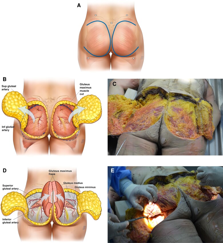
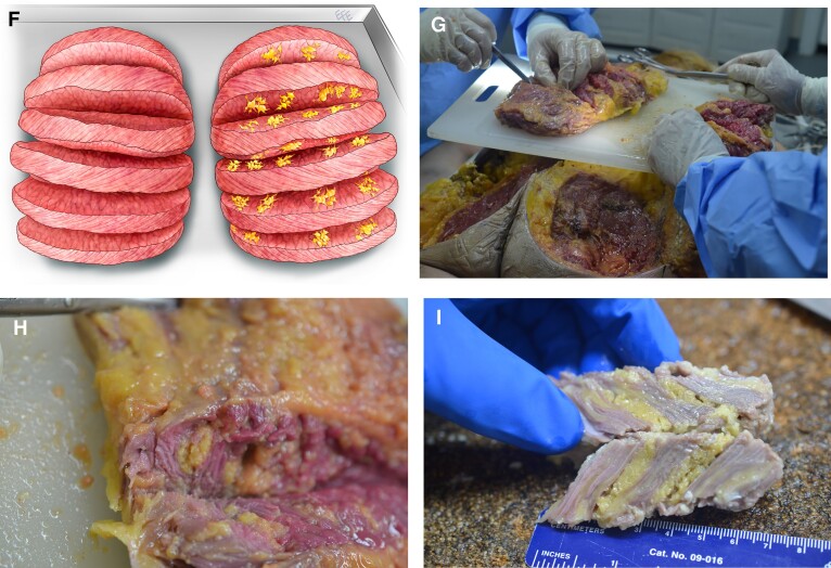
Figure 4.
Gluteal butterfly autopsy technique. (A) Step 1: initial skin incision of laterally based cutaneous flaps. (B) Step 2: the skin-subcutaneous flap is dissected from medial to lateral, exposing the deep gluteal fascia and the muscle belly of the gluteus maximus underneath the fascia. (C) The exposed deep gluteal fascia is seen at autopsy. (D) Step 3: the gluteus maximus is detached from lateral attachments and trochanter and reflected medially, exposing the gluteal arteries and veins. (E) Underbelly of gluteus maximus and exposed gluteal vessels noted at autopsy. (F) Step 4: the gluteus maximus muscle belly is cut horizontally in a bread-loaf fashion to document the fat graft position within the muscle belly. (G) Horizontal breadloaf incisions through the gluteus maximus muscle belly are shown at autopsy. (H, I) Close-up views of incised muscle belly with intramuscular fat grafts oriented in tracts within the multiple cannula tunnels. Artwork created by and published with permission from Dr Levent Efe, CMI.


