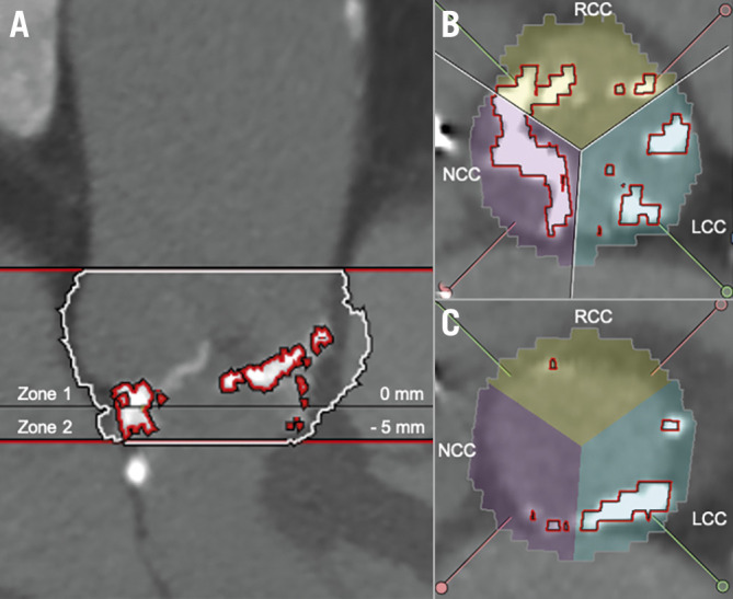
Figure 1. LVOT calcification quantification based on contrast-enhanced MSCT.

A) Aortic valve (AV) complex calcification quantification: Zone 1 (=annular plane, basal plane to the coronary ostia) and Zone 2 (=LVOT, basal plane to 5 mm deep in the LVOT). B) Distribution of calcification in Zone 1 (annular plane) according to AV leaflets in annular plane. C) Distribution of calcification in Zone 2 (LVOT) according to AV leaflets. LCC: left coronary cusp; NCC: non-coronary cusp; RCC: right coronary cusp</p>
