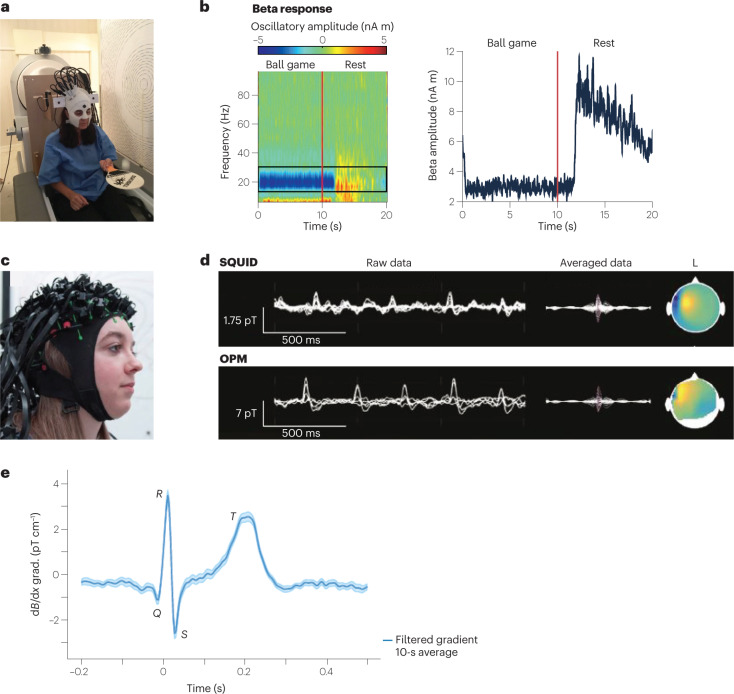
Fig. 3. Optically pumped magnetometer (OPM)-based magnetoencephalography (MEG).
a, Prototype wearable OPM-MEG. The subject can move their head during the measurement, as exemplified by the subject bouncing a tennis ball off a bat. b, Beta band oscillations recorded as a frequency spectrogram (left) and amplitude (right) during ball game and rest. c, Flexible wearable OPM-MEG helmet with 63 sensor mounts. d, SQUID-MEG (top) and OPM-MEG (bottom) recording of 11-year-old patient with refractory focal epilepsy. Left: superimposed data of multiple sensors showing filtered background brain activity and interictal epileptiform discharges (IEDs). Right: averaged IED data and magnetic field topography at the spike peak. e, OPM-magnetocardiography (MCG) measured at ambient conditions with a 87Rb magnetic gradiometer. Parts a,b adapted with permission from ref. 4, Springer Nature Ltd. Part c adapted with permission from ref. 149 under a Creative Commons licence CC BY 4.0. Part d adapted with permission from ref. 53, RSNA. Part e adapted with permission from ref. 51, APS.

