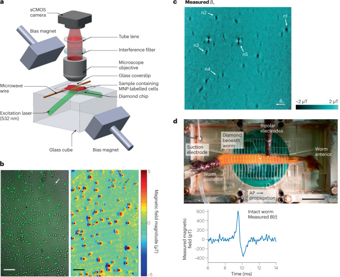
Fig. 4. Nitrogen–vacancy (NV)-centre-based magnetic sensing of biological samples.
a, Wide-field NV-diamond microscope for magnetic imaging of cells. sCMOS, scientific complementary metal–oxide–semiconductor. b, Wide-field imaging of biomarkers. Left: bright-field image overlayed with fluorescence image of SKBR3 cancer cells labelled with magnetic nanoparticles (MNPs) and stained with fluorescence dyes. Right: same field of view showing NV magnetic imaging of MNP-labelled cells. Scale bar, 100 µm. c, Magnetic field image of natural haemozoin crystals acquired with a NV-diamond microscope. Field of view is 39 × 39 µm². d, Top: image of NV-diamond set-up for single-neuron action potential (AP) magnetic measurement of a living specimen of Myxicola infundibulum (worm). Bottom: time trace of the magnetic field signal coming from a single-neuron action potential of M. infundibulum detected with the NV-diamond set-up. Part a adapted with permission from ref. 62, Springer Nature Ltd. Part b adapted with permission from ref. 63, Springer Nature Ltd. Part c adapted with permission from ref. 64 under a Creative Commons licence CC BY 4.0. Part d adapted with permission from ref. 66, PNAS.

