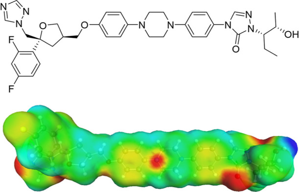
Figure 2.

Top: Posaconazole molecular structure. Bottom: Posaconazole σ-surface reported from COSMOquick; areas with positive polarization charge density are highlighted in red (H-bond acceptor), while areas with negative polarization charge density area are highlighted in blue (H-bond donor).
