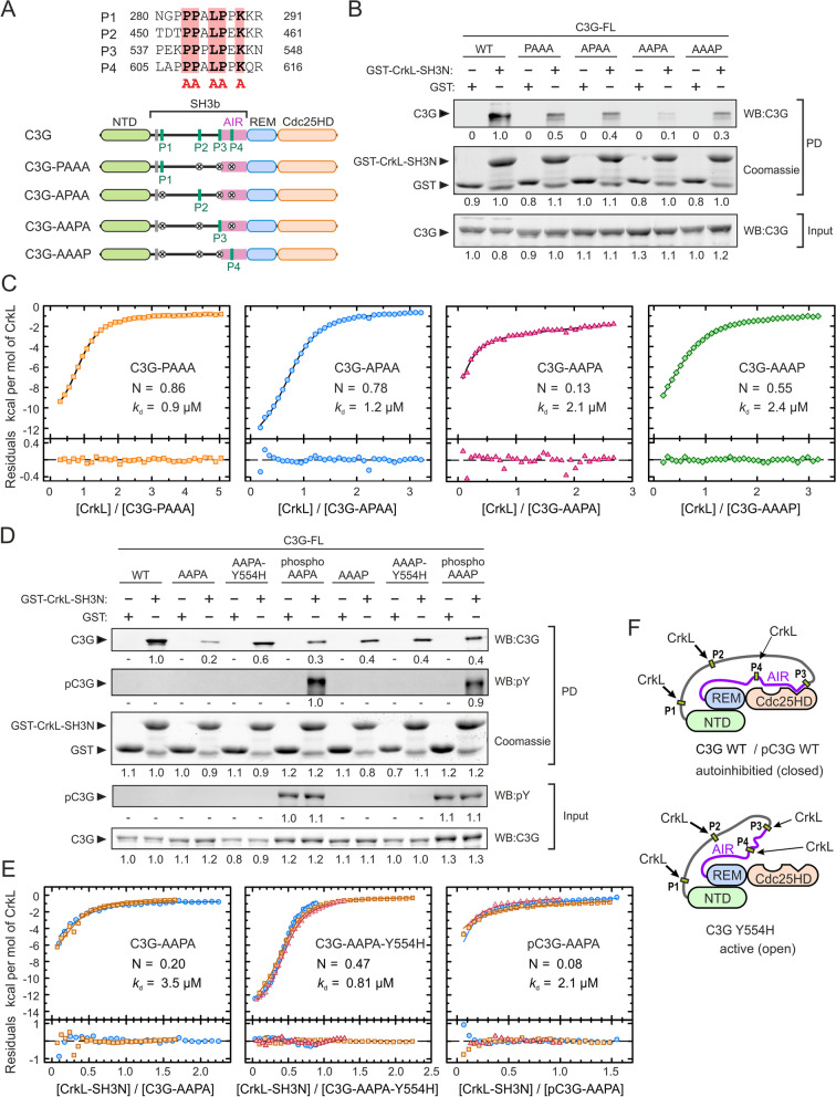
Fig. 2.
Binding of CrkL to individual sites in full-length C3G. A Sequences of the four PRMs of human C3G that are binding sites for CrkL. The five key residues in each motif that were mutated to Ala to disrupt CrkL binding are marked by red boxes. Schematic representation of the C3G mutants, each containing a single wild type PRM. B Analysis by pull-down (PD) of the interaction of GST-CrkL-SH3N with C3G full-length (FL) wild type (WT) and mutants. C3G was detected in the PD and input samples by western blot (WB). GST-CrkL-SH3N and GST (used as control) were visualized by coomassie staining. Numbers are the relative quantification within each panel. C ITC analysis of the binding of full-length CrkL to single-PRM mutants of C3G. The binding isotherms (1:1 binding models) and the residuals from the fitted model are shown. N is the estimated competent fraction that reflects the fraction of sites accessible for binding. The corresponding thermograms are shown in Additional file 1: Fig. S3A. D PD analysis of the binding of GST-CrkL-SH3N to C3G WT and single-PRM mutants, in combination with the activating mutation Y554H or after phosphorylation by Src. Numbers are relative quantification of bands as in (B). E ITC binding isotherms of the interaction of the SH3N domain of CrkL to C3G-AAPA, C3G-AAPA-Y554H, and Src-phosphorylated pC3G-AAPA. Thermograms are shown in Additional file 1; Fig. S3B. F Schematic models of the structure of autoinhibited C3G WT and the constitutive active mutant C3G Y554H. The P3 is exposed in the active conformation

