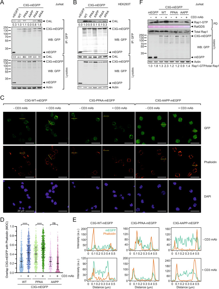
Fig. 5.
Role of the PRMs in the recruitment of C3G to the plasma membrane and the activation of Rap1 in Jurkat cells. A Analysis by co-immunoprecipitation (coIP) in Jurkat cells of the interaction between stably expressed exogenous C3G-mEGFP variants and endogenous CrkL. Proteins were immunoprecipitated with affinity resin against GFP. CrkL and mEGFP-tagged proteins were detected in cell lysates and in the IP by western blot (WB). B Analysis of the interaction between C3G-mEGFP and CrkL in HEK293T cells. mEGFP-tagged C3G WT and mutants were transiently expressed and the interaction with endogenous CrkL was analyzed by coIP as in (A). C Imaging of C3G-mEGFP (upper panels), and cortical actin (stained with phalloidin-iFluor 647, middle panels) in Jurkat cells expressing C3G wild type or the indicated mutants. Nuclei were stained with DAPI. Cells were plated on coverslips treated with poly-l-lysine and were unstimulated (anti-CD3) or stimulated with OKT-3 antibody against CD3. Representative fields are shown, in which the median of the Manders’ overlap coefficient (MOC) for the cells in each field is similar to the value observed for the total of cells analyzed. Scale bars, 20 μm. D Quantification of the co-localization of C3G-mEGFP with phalloidin-stained actin in confocal microscopy images of Jurkat cells as shown in (C). MOC values are shown as scatter plots (from left to right, n = 637, 466, 346, 565, 423, and 459 cells). Middle bars mark the median and whiskers are the 25th and 75th percentiles. Statistical analysis were done with non-parametric Kruskal–Wallis and Dunn´s multiple comparisons tests (****P < 0.0001, n.s. P > 0.05). E Fluorescence intensity of C3G-mEGFP and phalloidin staining along the yellow dashed lines in panel (C), which run across representative cells. F Analysis of Rap1 activation in Jurkat cells stably expressing C3G-mEGFP WT and mutants, or isolated mEGFP as control, before and after stimulation with antibody against CD3

