Fig. 3.
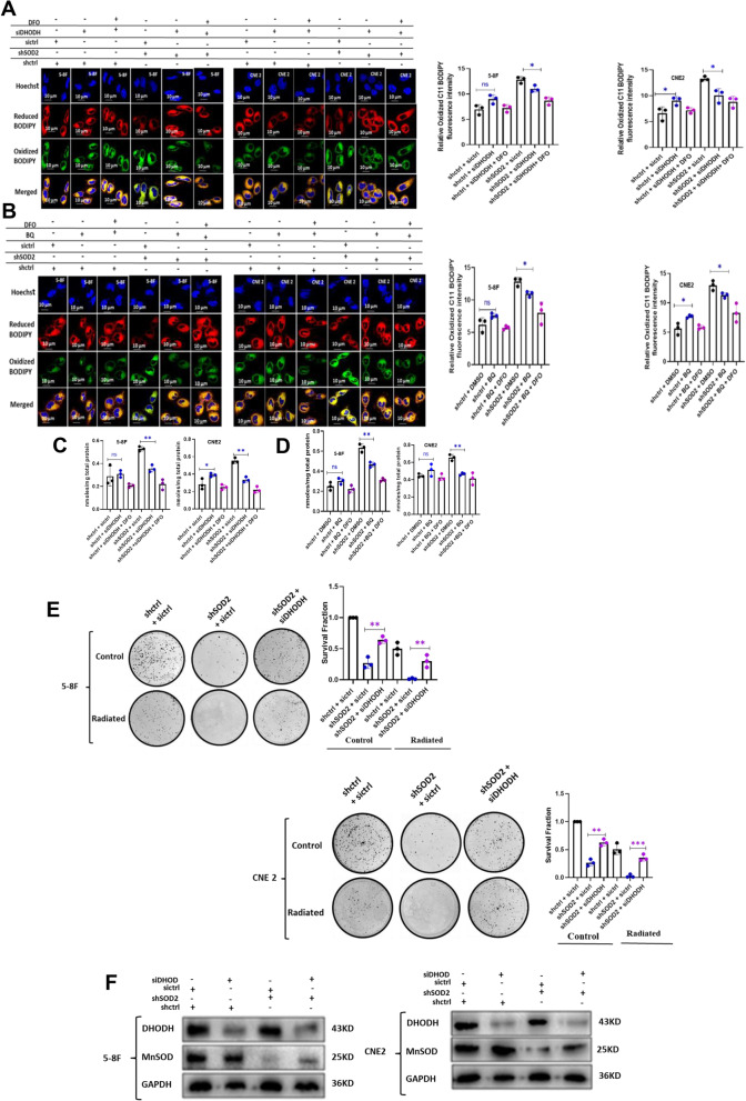
DHODH inhibition suppressed ferroptosis in SOD2 depleted cells. Confocal microscopy images and bar plots showing the fluorescence intensity of oxidized 581/591 C11 BODIPY in (A) SOD2 and DHODH double knockdown cells and (B) SOD2 knockdown cells treated with 5 µM BQ. DFO was further used to inhibit ferroptosis in DHODH inhibited and SOD2 knockdown, DHODH inhibited cells. MDA was down regulated in SOD2 depleted cells treated with (C) 5 nM siDHODH and (D) 5 µM BQ. E Restoration of colony formation by DHODH knockdown in SOD2 depleted 5-8F and CNE 2 cells. F Western blot showing DHODH and SOD2 double knockdown in 5-8F and CNE2 cells. The blots are from different parts of the same gel. n = 3 independent repeats. *P < 0.05; **P < 0.01. Radiation dosage: 4 Gy

