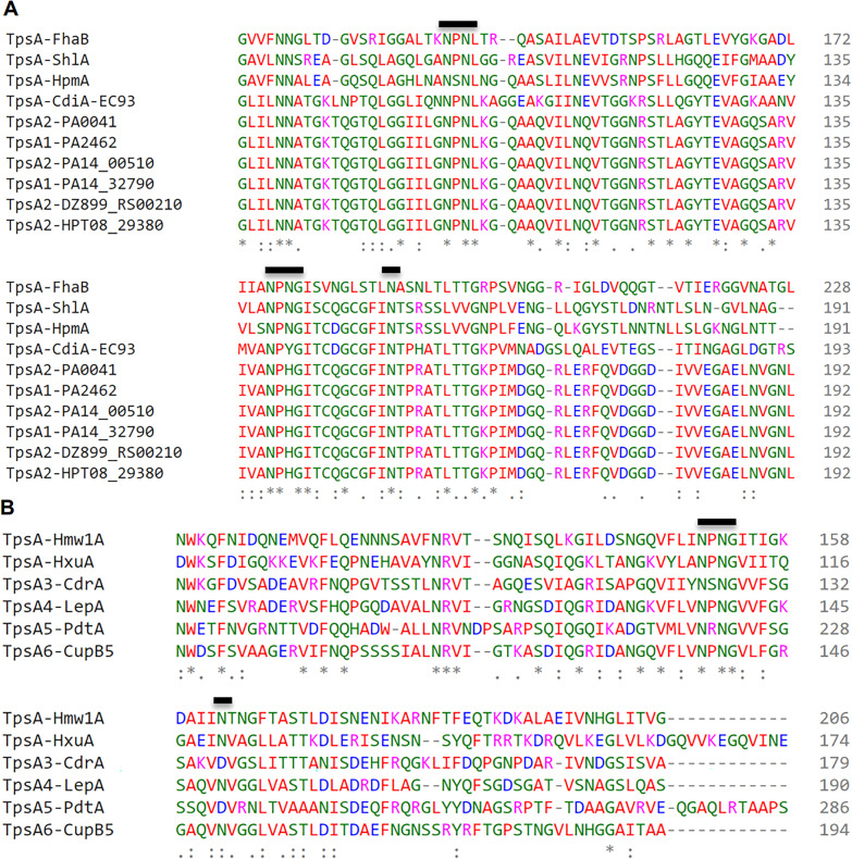
Fig. 2.
Multiple sequence alignment of the N-terminal region (1–360 aa) of representative TpsA proteins using CLUSTAL omega (https://www.ebi.ac.uk/Tools/msa/clustalo/). The numbering on the left corresponds to the amino acid number for each protein. N terminal amino acid residues of TpsA proteins (TpsA1 to 6) of P. aeruginosa were compared with, B. pertussis FHA protein (A) and H. influenzae HMW1 protein (B). Bold line indicates the position of motifs that have been identified in N-terminal TPS domains that are important for the secretion of TpsA proteins

