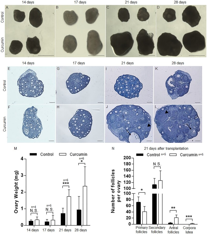
Fig. 2.
Increased ovarian and follicular development in vivo after curcumin treatment in vitro. Paired PD7 ovaries were treated with or without curcumin (100 µM) for 6 d followed by transplantation under the kidney capsules of ovariectomized recipient mice. Transplanted ovarian grafts were collected 14 to 28 d later. (A–D) Isolated paired ovaries for curcumin-treated versus controls after transplantation for 14, 17, 21, and 28 d. Scale bars: 1 mm. (E–L) The morphology of curcumin-treated (F, H, J, and L) and control (E, G, I, and K) ovaries after 14 to 28 d of engraftment (arrowheads, corpora lutea; arrows, antral follicles). Hematoxylin staining indicates nuclei. Scale bars: 50 µm. (M) The weight of the ovaries was measured 14 to 28 d after transplantation. (N) Distribution of follicles in ovaries treated with or without curcumin after transplantation for 21 d. Data are shown as the mean ± SD. *P < 0.05; **P < 0.01; ***P < 0.001 versus control.

