Fig. 5.

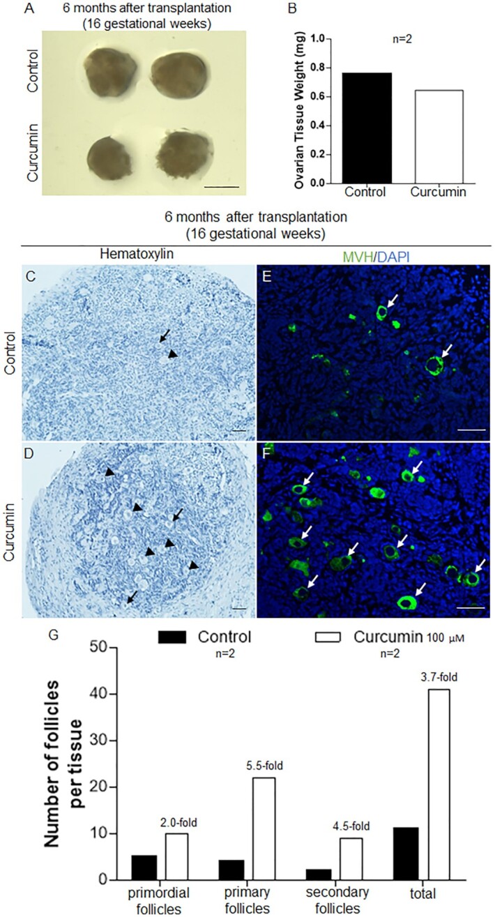
Curcumin treatment enhanced the survival and development of human follicles in the 16 gestational weeks fetal ovarian tissues. Ovarian fragments (16 gestational weeks) were carefully cut into small cubes, treated with or without curcumin (100 µM) for 6 d, and transplanted under the kidney capsule of ovariectomized SCID mice. (A) Isolated ovarian tissues for curcumin-treated versus controls after transplantation for 6 months. Scale bars: 1 mm. (B) The weight of the ovarian tissues was measured 6 months after transplantation. (C and D) The morphology of curcumin-treated (D) and control (C) ovarian tissues after 6 months of engraftment (arrowheads, growing follicles; arrows, primordial follicles). Hematoxylin staining indicates nuclei. Scale bars: 50 µm. (E and F) Immunofluorescent labeling of germ cells showing oocytes in the control (E) and curcumin-treated (F) tissues (arrows, oocytes; green: MVH, blue: DAPI). Scale bars: 50 µm. (G) Distribution of follicles in ovarian tissues treated with or without curcumin after transplantation for 6 months.
