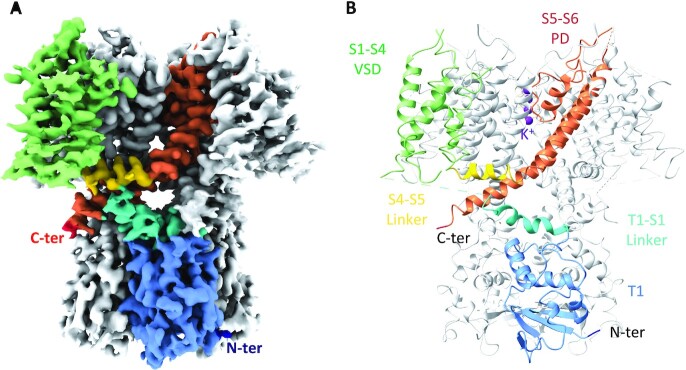
Fig. 1.
Structure of apo flWT-Kv3.1a. (A) Cryo-EM reconstruction of apo flWT-Kv3.1a. (B) Atomic model of apo flWT-Kv3.1a. The color code is identical in both figures and highlights the different domains and their organization in flWT-Kv3.1a. For clarity, only one monomer has been colored, the remaining three monomers are shown in light gray. The N-terminus end is colored in dark blue, the T1 domain in light blue, the T1-S1 linker in cyan, the VSD in light green, the S4–S5 linker in yellow, the PD in orange, and the C-terminus end in red. Potassium ions visible in the reconstruction were modeled and are colored in purple.

