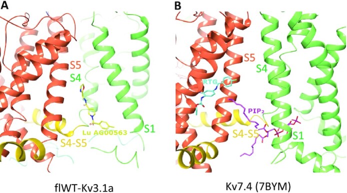
Fig. 4.

Comparison between the binding site of Lu AG00563 in flWT-Kv3.1a and the binding sites of PIP2 and RTG in Kv7.4. (A) Binding site discovered for Lu AG00563 at the interface between S1, S4, and the S4–S5 linker. The S1 and S4 helices are shown in light green, S5 in orange, and the S4–S5 linker in yellow. Lu AG00563 is shown in yellow. (B) Comparison with the Kv7.4 structure (pdb code: 7bym), highlighting similarities with the PIP2 binding site showing interactions with S1, S4, and the S4–S5 linker. In contrast, the RTG binding site is located next to the pore (S5) domain. PIP2 is shown in purple and RTG is shown in Cyan.
