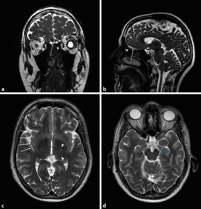
Fig. 1.
a Magnetic resonance imaging (MRI) of the skull; coronal T2-weighted image. Olfactory bulb (arrow), olfactory sulcus (triangles), olfactory mucosa (star). b MRI of the skull; sagittal T2-weighted image. Olfactory bulb (arrow), olfactory mucosa (star). c MRI of the skull; axial T2-weighted image. Insula (arrows), thalamus (triangles). d MRI of the skull; axial T2-weighted image. Amygdala (white circle), hippocampus (blue circle)

