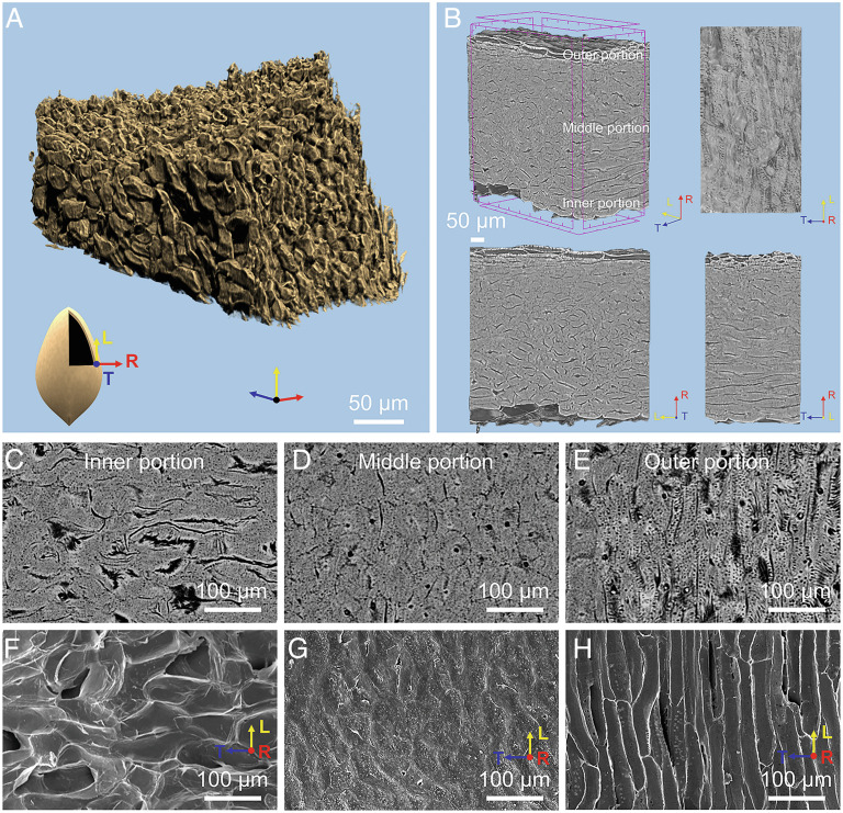
Fig. 2.
X-ray micro-CT and microscopy characterization of seed shells. (A) 3D reconstruction of the seed shell derived from synchrotron X-ray micro-CT showing the packed sclereids. (B) The micro-tomography images display the three directions: longitudinal (L), tangential (T), and radial (R). (C–E) The micro-tomography images of the inner portion of the seed shell, middle portion, and the outer portion (slightly aligned along the T direction), and (F–H) the corresponding SEM images of the three areas.

