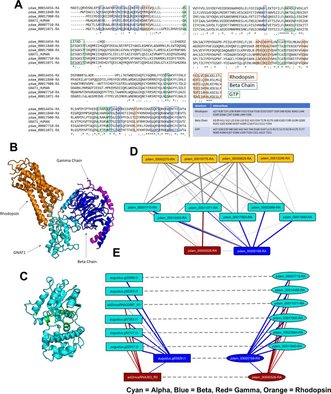
Fig 5. Identification of putative G protein subunits as candidates of immediate downstream signaling partners for GPCRs.
(A) MSA of GNAT1 with coral homologues and crystal structures of GNAT1. Amino acids of GNAT1 which interact with rhodopsin, GNB1 (beta chain), and GTP are boxed in blue, red, and green, respectively. The same amino acids are also listed in the table. In the crystal structures, GNAT1 (cyan) binding interfaces with rhodopsin (orange) and the beta chain (blue) are shown in (B) and the GTP binding pocket (green) is shown in (C). (D) Predicted interactions in P. damicornis between alpha (cyan), beta (blue), gamma (orange) chains and rhodopsin (orange). (E) Predicted candidate G protein interactions in P. damicornis, and the predicted interactions of the corresponding candidates in M. capitata (solid line: Predicted interaction; dashed line: Best-bidirectional BLAST hit).

