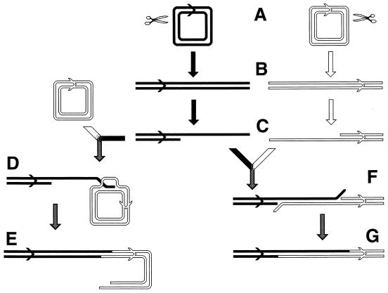
FIG. 32.
Comparison of the double-strand end invasion with single-strand annealing reactions. Duplex DNA is shown as double lines (open or solid); the cos (packaging) site of λ is shown as a chevron. (A) Circular λ chromosomes. (B) The chromosomes in panel A are cut at different and unique sites in vivo (marked with scissors in panel A. (C) The ends are processed by λ exo to generate 3′ overhangs. (D and E) The RecA-catalyzed invasion of the 3′ overhang into an intact λ chromosome with the following resolution of the joint molecule (190). (F) Alternatively, the 3′ overhang is annealed by Beta with a complementary 3′ overhang of another linear λ chromosome, cut at a different location. (G) Completion of the SSA recombination. Note that the hybrid region in panel E is expected to be shorter than in panel G.

