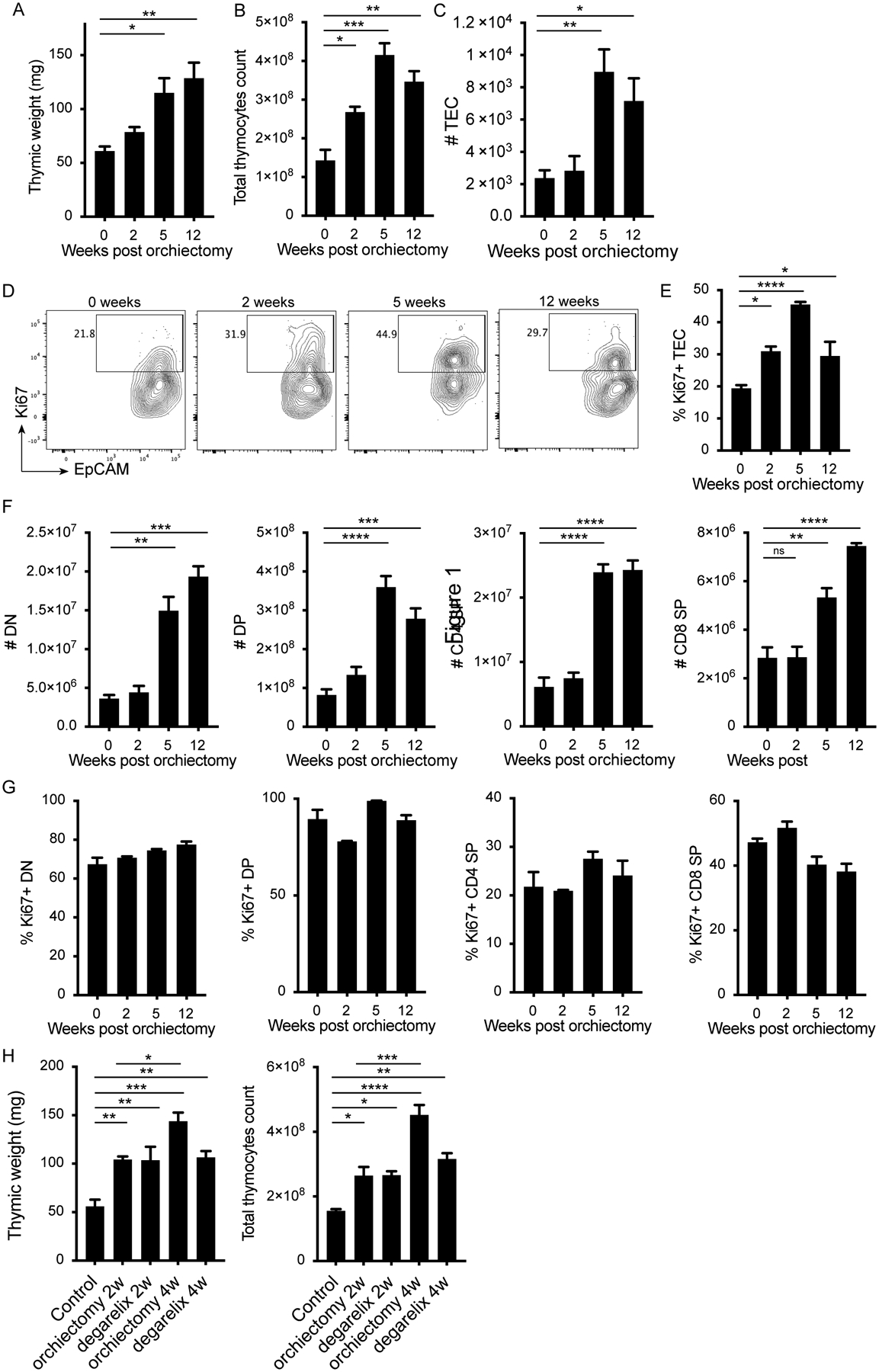
Figure 1. ADT promotes thymic regeneration through proliferation of TEC but not thymocytes.

12-week old male mice were orchiectomized, and thymi were harvested at 2, 5 and 12 weeks later. A) Thymic weight, B) total thymocyte count and C) number of thymic epithelial cells (TEC) at time of harvest. D) Representative flow plots gated on TECs (CD45−Epcam+) and showing EpCAM and Ki67 expression, and E) quantification of percent Ki67+ TEC at 0, 2, 5 and 12 weeks post orchiectomy. F) Numbers and G) percent Ki67+ of DN, DP, CD4 SP and CD8 SP in the thymus at the indicated times. H) 12 week old male mice were orchiectomized or chemically castrated using degarelix, and thymi were harvested 2 or 4 weeks later. Graphs show thymic weight and total thymocyte counts after the indicated treatments. 3 animals per group. Data representative of 3 experiments. One-way ANOVA with Tukey multiple comparison, * P<0.05, ** P<0.01, *** P<0.001, **** P<0.0001.
