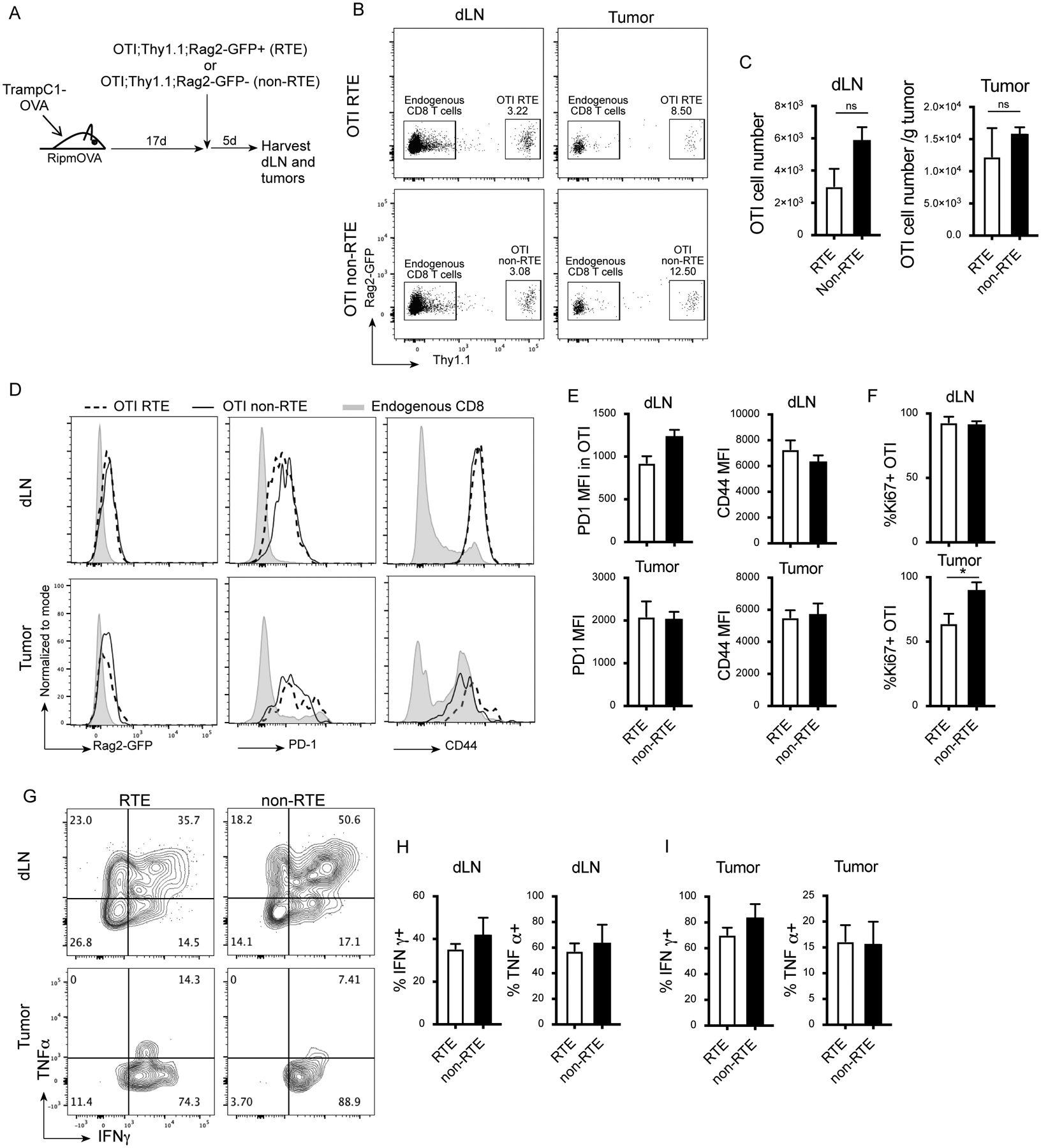
Figure 3. Tumor-specific RTE contribute to the anti-tumor immune response.

A) Experimental design. B) Representative flow plots (gated on live, TCRβ+) showing Rag2-GFP and Thy1.1 staining in the dLN and tumor of animals that received adoptive transfer of RTE OTI or non-RTE OTI. C) Cell number of recovered OTI cells in the dLN and tumor. D) Representative histograms (gated on live, TCRβ+, CD8+, Thy1.1+ or Thy1.1-) showing Rag2-GFP, PD1 and CD44 expression in the OTI cells and host endogenous CD8 T cells. E) Quantification of PD1 and CD44 MFI in OTI the dLN and tumor. F) Percent Ki67+ OTI cells. G-I) dLN cells and tumor infiltrating lymphocytes (TIL) were stimulated in vitro with SIINFEKL peptide and stained for intracellular cytokines. G) Representative flow plots (gated on live, TCRβ+, CD8, Thy1.1+) showing IFNγ and TNFα expression adoptively transferred RTE and non-RTE in the dLN and tumor. H-I) Quantification of percent IFNγ+ and TNFα+ among adoptively transferred RTE and non-RTE in the dLN (H) and tumor (I). 3 animals per group. Data are representative of 2 experiments. Unpaired two-tailed Student t test, * P<0.05.
